Review
. 2022 Feb 27;23(5):2617.
doi: 10.3390/ijms23052617.

The Pathogenesis of Cardiac Fibrosis: A Review of Recent Progress

Affiliations
Review

The Pathogenesis of Cardiac Fibrosis: A Review of Recent Progress

Kazuaki Maruyama et al. Int J Mol Sci. .

Abstract

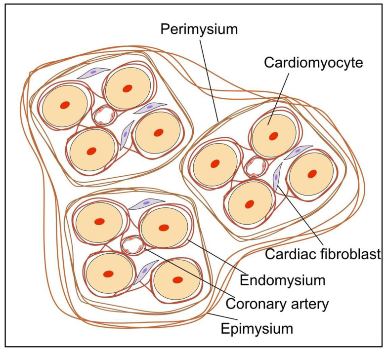
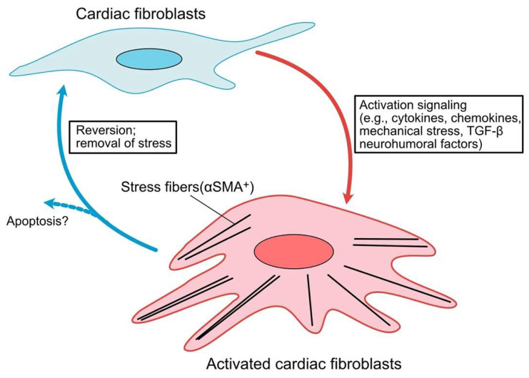
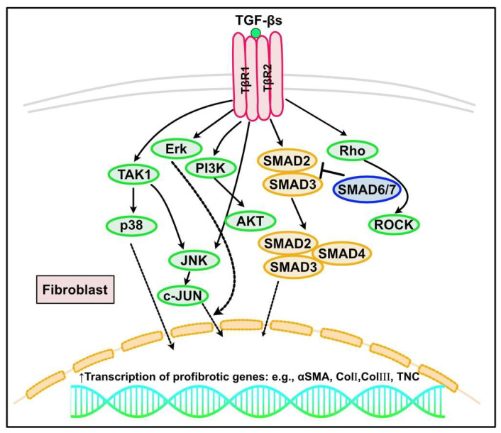
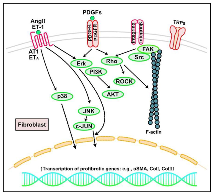
Fibrosis is defined as the excessive deposition of extracellular matrix (ECM) proteins in the interstitium. It is an essential pathological response to chronic inflammation. ECM protein deposition is initially protective and is critical for wound healing and tissue regeneration. However, pathological cardiac remodeling in excessive and continuous tissue damage with subsequent ECM deposition results in a distorted organ architecture and significantly impacts cardiac function. In this review, we summarized and discussed the histologic features of cardiac fibrosis with the signaling factors that control it. We evaluated the origin and characteristic markers of cardiac fibroblasts. We also discussed lymphatic vessels, which have become more important in recent years to improve cardiac fibrosis.

Keywords: cardiac fibrosis; marker of cardiac fibroblast; pathogenesis of cardiac fibrosis; signaling of cardiac fibrosis.


Conflict of interest statement

The authors declare no conflict of interest.

Figures

Figure 1
Figure 1
Schematic image of the cardiac interstitial collagen network. The endomysium surrounds and interconnects individual cardiomyocytes. The perimysial weave segregates cardiomyocytes into groups. The epimysium surrounds and clusters large numbers of myofibres.
Figure 2
Figure 2
Progression and characterization of a cardiac fibroblast to myofibroblast conversion. In response to cardiac injury, cytokines, chemokines and neurohumoral factors, resident cardiac fibroblasts (CFs) become activated with increasing expression of α smooth muscle actin (α-SMA). These activated CFs may undergo apoptosis following the scar formation. Activated CFs are capable of de-differentiating upon removal of stress stimuli.
Figure 3
Figure 3
TGF-β signaling in cardiac fibrosis. TGF-β could induce the signal transduction via the canonical (SMAD-dependent) and non-canonical (SMAD-independent) pathways. In the canonical pathway, TGF-β1 binds to and causes heterodimerization of TGF-β receptor type 1 (TβRI, also known as activin-like kinase (ALK) 5) and the type II receptor (TβRII), leading to the phosphorylation of SMAD2/SMAD3, which subsequently form a complex with SMAD4 and translocate into the nucleus, acting as a transcriptional factor to regulate the fibrotic gene expression (e.g., αSMA, collagen I, III or TNC). SMAD6/7 are inhibitory SMADs to inhibit transcription of SMAD2 and SMAD3. In canonical pathways, TGF-β1 can also induce SMAD-independent noncanonical signaling that involves several mitogen-activated protein kinases, including extracellular signal-regulated kinase (Erk), c-Jun-N-terminal kinase (JNK), TGF-β-activated kinase 1 (TAK1), Rho family of small GTPase, and p38 MAPK pathways.
Figure 4
Figure 4
Other signaling pathways regulating cardiac fibrosis. In response to increased mechanical stress and cardiac injury, inflammatory signaling, growth factors (e.g., platelet-derived growth factors (PDGFs)), neurohumoral pathways (e.g., angiotensin (Ang) II, endothelin (ET)-1), and mechanosensitive pathways mediated by integrins and ion channels such as transient receptor potential cation channels (TRPs) can activate fibroblasts into myofibroblasts, leading to excess extracellular matrix protein deposition and cardiac fibrosis. AT1, angiotensin type 1 receptor; PDGFR, PDGF receptor; ERK, extracellular signal regulated kinase; PI3K, phosphoinositide 3-kinase; JNK, c-JUN N-terminal kinase; αSMA, α-smooth muscle actin; ROCK, Rho-associated protein kinases; FAK, focal adhesion kinase.
Figure 5
Figure 5
Canonical WNT/β-catenin signaling in cardiac fibrosis. In the absence of Wnt ligands, cytosolic β-catenin is degraded by the destruction complex, which includes Axin and adenomatous polyposis coli (APC), glycogen synthase kinase (GSK)-3β and casein kinase (CK)1, protein phosphatase 2A (PP2A), and β-transducin repeat-containing protein (β-TrCP). After a Wnt ligand binds to the receptor Frizzled (Fz) and the receptor-related protein 5 or 6 (LRP5/6) coreceptor, the Wnt–Fz–LRP5/6 complex recruits Disheveled (DVL) and Axin through the intracellular domains of Fz and LRP5/6, resulting in β-catenin stabilization. The increased nuclear levels of β-catenin promote interaction with T cell factor/lymphoid enhancer factor (TCF/LEF) transcription factor to regulate Wnt-responsive fibrotic genes.

References

    1. Rossignol P., Hernandez A.F., Solomon S.D., Zannad F. Heart failure drug treatment. Lancet. 2019;393:1034–1044. doi: 10.1016/S0140-6736(18)31808-7. - DOI - PubMed
    1. Frangogiannis N.G. Cardiac fibrosis. Cardiovasc. Res. 2020;117:1450–1488. doi: 10.1093/cvr/cvaa324. - DOI - PMC - PubMed
    1. Sainio A., Järveläinen H. Extracellular matrix-cell interactions: Focus on therapeutic applications. Cell Signal. 2020;66:109487. doi: 10.1016/j.cellsig.2019.109487. - DOI - PubMed
    1. Li L., Zhao Q., Kong W. Extracellular matrix remodeling and cardiac fibrosis. Matrix Biol. 2018;68:490–506. doi: 10.1016/j.matbio.2018.01.013. - DOI - PubMed
    1. Krejci J., Mlejnek D., Sochorova D., Nemec P. Inflammatory Cardiomyopathy: A Current View on the Pathophysiology, Diagnosis, and Treatment. Biomed. Res. Int. 2016;2016:4087632. doi: 10.1155/2016/4087632. - DOI - PMC - PubMed

Substances

LinkOut - more resources