. 2022 Mar 1;23(5):2729.
doi: 10.3390/ijms23052729.

Mechanism Underlying the Bypass of Apurinic/Pyrimidinic Site Analogs by Sulfolobus acidocaldarius DNA Polymerase IV

Affiliations

Mechanism Underlying the Bypass of Apurinic/Pyrimidinic Site Analogs by Sulfolobus acidocaldarius DNA Polymerase IV

Qin-Ying Huang et al. Int J Mol Sci. .

Abstract

The spontaneous depurination of genomic DNA occurs frequently and generates apurinic/pyrimidinic (AP) site damage that is mutagenic or lethal to cells. Error-prone DNA polymerases are specifically responsible for the translesion synthesis (TLS) of specific DNA damage, such as AP site damage, generally with relatively low fidelity. The Y-family DNA polymerases are the main error-prone DNA polymerases, and they employ three mechanisms to perform TLS, including template-skipping, dNTP-stabilized misalignment, and misincorporation-misalignment. The bypass mechanism of the dinB homolog (Dbh), an archaeal Y-family DNA polymerase from Sulfolobus acidocaldarius, is unclear and needs to be confirmed. In this study, we show that the Dbh primarily uses template skipping accompanied by dNTP-stabilized misalignment to bypass AP site analogs, and the incorporation of the first nucleotide across the AP site is the most difficult. Furthermore, based on the reported crystal structures, we confirmed that three conserved residues (Y249, R333, and I295) in the little finger (LF) domain and residue K78 in the palm subdomain of the catalytic core domain are very important for TLS. These results deepen our understanding of how archaeal Y-family DNA polymerases deal with intracellular AP site damage and provide a biochemical basis for elucidating the intracellular function of these polymerases.

Keywords: AP site analogs; Dbh; Sulfolobus acidocaldarius; little finger domain; translesion synthesis.


Conflict of interest statement

The authors declare no conflict of interest.

Figures
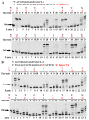
Figure 1
Figure 1
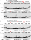
Primer extension of paired or mispaired 3′ base pairs with a bulging spacer. The PT DNA substrates are shown at the top of the figure, where the letter X denotes spacers C3 (A) and C6 (B). The first 3′ base of the primer strand is A, T, C, or G. The first base upstream of the AP site in the template strand is A, T, C, or G. Four primer strands were annealed with four different template strands to form 16 kinds of PT DNA substrates with a bulging spacer C3 or C6, which were incubated with 0.5 µM Dbh and dNTPs at 45 °C for 0, 10, 20, and 60 min. Asterisks denote the terminal fluorescent group FAM.
Figure 2
Figure 2
Single dNMP incorporation during AP site bypass. The PT DNA substrates are shown at the top of the figure, where the letter X denotes spacers C3 (A) and C6 (B). The first base upstream of the AP site in the template strand is A, T, C, or G. The PT DNA substrates were incubated with 0.1 µM Dbh in the presence of dATP, dCTP, dGTP or dTTP at 45 °C for 0, 10, 20, and 60 min. Asterisks denote the terminal fluorescent group FAM.
Figure 3
Figure 3
Primer extension with increasing pairing numbers of 3′ base pairs with a bulging spacer. The PT DNA substrates are shown at the top of the figure. The letter X denotes a skipped base A and spacers C3, C6, C12, S9, S18, and dS. The symbol X denotes the normal DNA template. The PT DNAs, with 3′ base pairs of 1 bp (A), 2 bp, (B) and 3 bp (C) after the bulging spacer or base A, were used as substrates to characterize the extension reactions. The PT DNA substrates were incubated with 0.1 µM Dbh and dNTPs at 45 °C for 0, 1, 3, 10, 30, and 60 min. Asterisks denote the terminal fluorescent group FAM.
Figure 4
Figure 4
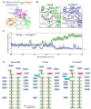
Effect of spacer C3 on TLS efficiency. (A) Schematic depiction of the domain composition of Dbh and its modeled structure with bound PT DNA carrying a free spacer C3. (B) Modeling of the formation of the phosphodiester bond between the incoming dNTP and the 3′-OH of the primer paired with the DNA template strand with a free (left panel) or bulging (right panel) spacer C3. (C) Distance between the 3′-OH of the primer strand and the alpha phosphate group of the incoming dNTP. PT DNA with a free (C3-0 bp) or bulging (C3-1 bp) spacer C3 was used to calculate the distance. (D) Hydrogen bond interactions between Dbh and normal PT DNA substrate (left panel) or the counterparts carrying a spacer C3 in the DNA template strand (the middle panel is for free spacer C3, and the right panel is for bulging spacer C3).
Figure 5
Figure 5
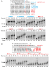
Effect of the bulging loop on TLS. The PT DNA substrates are shown at the top of the figure, where the letter X denotes bases A, T, C, G, and dSpacer, and the letter Y denotes bases A, C, and dSpacer. The symbol X denotes without spacer or base. (A) Effect of the base mismatches (T:C mismatch and T:dS mismatch) and a bulging base loop or dSpacer loop (1-mer loops of dS, C, G, T, and A) on TLS efficiency. (B) Effect of the size of the bulging loop (dS, dS-C, dS-C2, and dS-C3) on TLS efficiency. The PT DNA substrates were incubated with 0.1 µM Dbh and dNTPs at 45 °C for 0, 1, 3, 10, 30, and 60 min. Asterisks denote the terminal fluorescent group FAM.
Figure 6
Figure 6
TLS by LF domain mutants of Dbh. The PT DNA substrates are shown at the top of the figure, where the letter X denotes base C and spacers C3 and C6. The symbol X denotes the normal DNA template. The PT DNA substrates, with 3′-pairing two normally paired base pairs downstream of the AP site in the template strand, were incubated with dNTPs and 0.2 µM wt Dbh (A), or the mutant Y249A (B), I287D (C), I289D (D), I295D (E), or R333A (F) at 45 °C for 0, 10, 20, and 60 min. 2 bp downstream of the 1-mer loop. Asterisks denote the terminal fluorescent group FAM.
Figure 7
Figure 7
TLS by palm and linker mutants of Dbh. The PT DNA substrates are shown at the top of the figure, where the letter X denotes base A (normal DNA template) and spacer C12. DNA substrates were incubated with dNTPs and 0.2 µM wt Dbh (A), or the mutant K78A (B), R283A (C), or K337A (D), at 45 °C for 0, 10, 20, and 60 min. Asterisks denote the terminal fluorescent group FAM.

Similar articles

References

    1. MacNeill S. Composition and Dynamics of the Eukaryotic Replisome: A Brief Overview. Subcell. Biochem. 2012;62:1–17. doi: 10.1007/978-94-007-4572-8_1. - DOI - PubMed
    1. Hübscher U., Maga G., Spadari S. Spadari, Eukaryotic DNA Polymerases. Annu. Rev. Biochem. 2002;71:133–163. doi: 10.1146/annurev.biochem.71.090501.150041. - DOI - PubMed
    1. O’Donnell M., Langston L., Stillman B. Principles and Concepts of DNA Replication in Bacteria, Archaea, and Eukarya. Cold Spring Harb. Perspect. Biol. 2013;5:a010108. doi: 10.1101/cshperspect.a010108. - DOI - PMC - PubMed
    1. Kelman L.M., Kelman Z. Archaeal DNA Replication. Annu. Rev. Genet. 2014;48:71–97. doi: 10.1146/annurev-genet-120213-092148. - DOI - PubMed
    1. Sarmiento F., Long F., Cann I., Whitman W.B. Diversity of the DNA Replication System in the Archaea Domain. Archaea. 2014;2014:675946. doi: 10.1155/2014/675946. - DOI - PMC - PubMed

Substances

LinkOut - more resources