. 2022 Mar 4;23(5):2828.
doi: 10.3390/ijms23052828.

Production and Stabilization of Specific Upregulated Long Noncoding RNA HOXD-AS2 in Glioblastomas Are Mediated by TFE3 and miR-661, Respectively

Affiliations

Production and Stabilization of Specific Upregulated Long Noncoding RNA HOXD-AS2 in Glioblastomas Are Mediated by TFE3 and miR-661, Respectively

Yiming Qin et al. Int J Mol Sci. .

Erratum in

Abstract

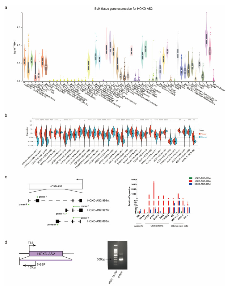
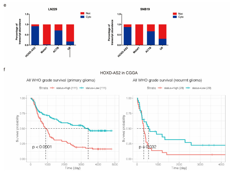
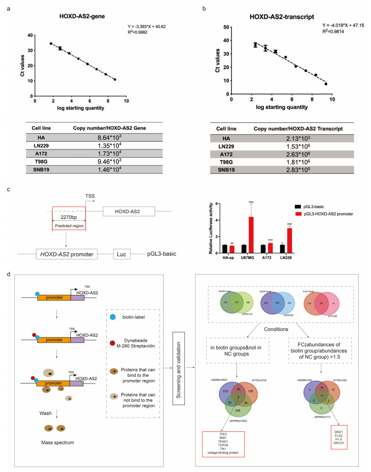
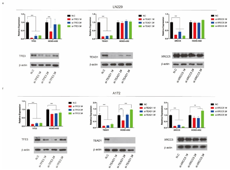
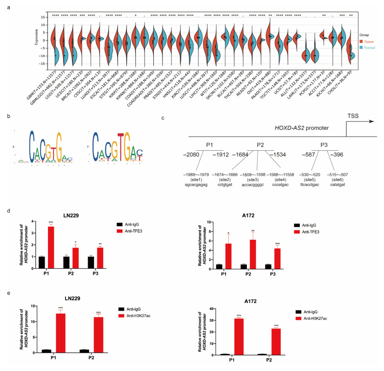
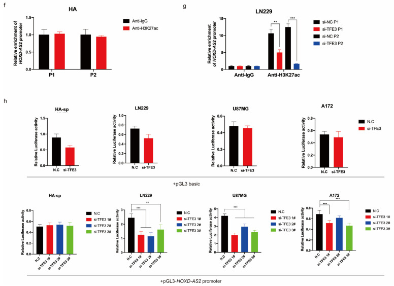
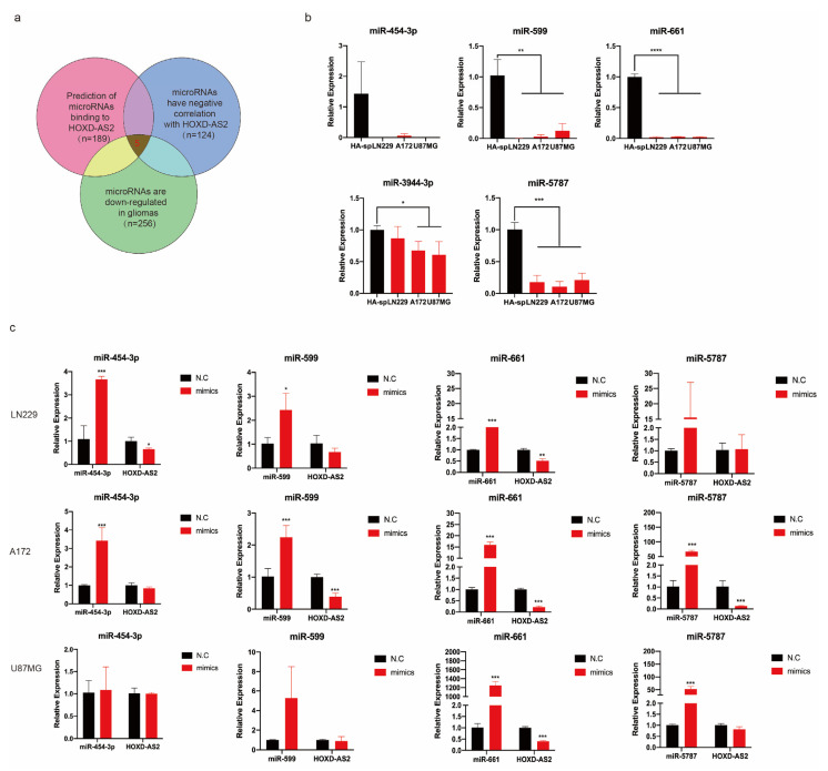
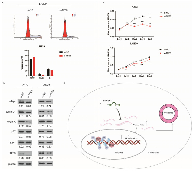
Differential expression of long noncoding RNAs (lncRNA) plays a key role in the development of gliomas. Because gliomas are the most common primary central nervous system tumor and glioblastomas have poor prognosis, it is urgent to develop new diagnostic methods. We have previously reported that lncRNA HOXD-AS2, which is specifically up-regulated in gliomas, can activate cell cycle and promote the development of gliomas. It is expected to be a new marker for molecular diagnosis of gliomas, but little is known about HOXD-AS2. Here, we demonstrate that TFE3 and miR-661 maintain the high expression level of HOXD-AS2 by regulating its production and degradation. We found that TFE3 acted as a transcription factor binding to the HOXD-AS2 promoter region and raised H3K27ac to activate HOXD-AS2. As the cytoplasmic-located lncRNA, HOXD-AS2 could be degraded by miR-661. This process was inhibited in gliomas due to the low expression of miR-661. Our study explains why HOXD-AS2 was specifically up-regulated in gliomas, helps to understand the molecular characteristics of gliomas, and provids insights for the search for specific markers in gliomas.

Keywords: HOXD-AS2; TFE3; glioma; miR-661.


Conflict of interest statement

The authors declare no conflict of interest.

Figures

Figure 1
Figure 1
HOXD-AS2 is up-regulated in gliomas and correlated with prognosis. (a) UCSC database showed the expression of HOXD-AS2 in normal tissues (http://genome.ucsc.edu, accessed on 23 January 2022). (b) The expression of HOXD-AS2 in different cancer types analyzed by TCGA and GTEx database. The p-values were calculated using a Wilcoxon test by R (version 4.0.5). Significant results were presented as * p < 0.05, ** p < 0.01, *** p < 0.001, **** p < 0.0001. (c) Pattern diagram of the specific primers for HOXD-AS2 three transcripts (left). Relative expression of HOXD-AS2 in astrocyte cell lines, glioblastoma cell lines and glioma stem cell lines were quantified by RT-qPCR (right). (d) HOXD-AS2 full-length identification by 5′RACE. (e) Detecting the expression of HOXD-AS2 in cytoplasm and nucleus by RT-qPCR. MALAT1, ACTB and U6 as positive controls. (f) Kaplan–Meier survival analysis of OS in GBM patients based on HOXD-AS2 expression in CGGA (Mantel–Cox test).
Figure 1
Figure 1
HOXD-AS2 is up-regulated in gliomas and correlated with prognosis. (a) UCSC database showed the expression of HOXD-AS2 in normal tissues (http://genome.ucsc.edu, accessed on 23 January 2022). (b) The expression of HOXD-AS2 in different cancer types analyzed by TCGA and GTEx database. The p-values were calculated using a Wilcoxon test by R (version 4.0.5). Significant results were presented as * p < 0.05, ** p < 0.01, *** p < 0.001, **** p < 0.0001. (c) Pattern diagram of the specific primers for HOXD-AS2 three transcripts (left). Relative expression of HOXD-AS2 in astrocyte cell lines, glioblastoma cell lines and glioma stem cell lines were quantified by RT-qPCR (right). (d) HOXD-AS2 full-length identification by 5′RACE. (e) Detecting the expression of HOXD-AS2 in cytoplasm and nucleus by RT-qPCR. MALAT1, ACTB and U6 as positive controls. (f) Kaplan–Meier survival analysis of OS in GBM patients based on HOXD-AS2 expression in CGGA (Mantel–Cox test).
Figure 2
Figure 2
Overexpression of HOXD-AS2 in glioblastomas was associated with transcriptional activation. (a,b) Absolute quantitative PCR in gene level (a) and transcription level (b). (c) The promoter region of HOXD-AS2 transcriptional activity was detected by luciferase report assay in astrocyte cell HA-sp and glioblastoma cell lines LN229, A172 and U87MG. (d) DNA-pull down/MS assay was used to find the factors which binding to the HOXD-AS2 promoter. Flow chart provided for screening of potential transcription factors regulating HOXD-AS2. (e,f) The expression of HOXD-AS2 was detected by RT-qPCR and Western blot after transfecting with TFE3/TEAD1/XRCC5 siRNAs in LN229 (e) and A172 (f). Data are presented as the mean ± SEM from three independent experiments. Significant results were presented as ** p < 0.01, *** p < 0.001. Two-tailed Student’s t-test was used in (d). (e,f) were analyzed by ANOVA.
Figure 2
Figure 2
Overexpression of HOXD-AS2 in glioblastomas was associated with transcriptional activation. (a,b) Absolute quantitative PCR in gene level (a) and transcription level (b). (c) The promoter region of HOXD-AS2 transcriptional activity was detected by luciferase report assay in astrocyte cell HA-sp and glioblastoma cell lines LN229, A172 and U87MG. (d) DNA-pull down/MS assay was used to find the factors which binding to the HOXD-AS2 promoter. Flow chart provided for screening of potential transcription factors regulating HOXD-AS2. (e,f) The expression of HOXD-AS2 was detected by RT-qPCR and Western blot after transfecting with TFE3/TEAD1/XRCC5 siRNAs in LN229 (e) and A172 (f). Data are presented as the mean ± SEM from three independent experiments. Significant results were presented as ** p < 0.01, *** p < 0.001. Two-tailed Student’s t-test was used in (d). (e,f) were analyzed by ANOVA.
Figure 3
Figure 3
TFE3 regulated the expression of HOXD-AS2. (a) TCGA and GTEx database demonstrated the expression of TFE3 in different tumors. The p-values were calculated using a Wilcoxon test by R (version 4.0.5). Significant results were presented as * p < 0.05, ** p < 0.01, *** p < 0.001, **** p < 0.0001. (b,c) The binding motifs of TFE3 (b) and binding sites on HOXD-AS2 promoter (c) were predicted by JASPAR (https://jaspar.genereg.net, accessed on 23 January 2022). (d) ChIP assays tested the enrichment of P1/P2/P3 fragments on the HOXD-AS2 promoter in glioma cells LN229 and A172; anti-IgG was used as the negative control group. (e,f) ChIP assays tested the enrichment of P1/P2 fragments on the HOXD-AS2 promoter in glioma cells, LN229 and A172 (e), astrocyte cell HA (f) and anti-IgG as the negative control group. (g) LN229 transfected with TFE3 siRNAs and performed ChIP-qPCR as above. (h) Luciferase activity assays were performed in astrocyte cell HA-sp and glioma cells included LN229, U87MG and A172, which were transfected with pGL3-basic vector or HOXD-AS2 promoter-containing pGL3 reporter vector and TFE3 siRNAs; firefly luciferase activity was detected and normalized by renilla luciferase activity. The data are shown as the means ± s.d. Two-tailed Student’s t-test was used in (dg). (h) was compared with control by ANOVA. Significant results were presented as * p < 0.05, ** p < 0.01, *** p < 0.001, **** p < 0.0001.
Figure 3
Figure 3
TFE3 regulated the expression of HOXD-AS2. (a) TCGA and GTEx database demonstrated the expression of TFE3 in different tumors. The p-values were calculated using a Wilcoxon test by R (version 4.0.5). Significant results were presented as * p < 0.05, ** p < 0.01, *** p < 0.001, **** p < 0.0001. (b,c) The binding motifs of TFE3 (b) and binding sites on HOXD-AS2 promoter (c) were predicted by JASPAR (https://jaspar.genereg.net, accessed on 23 January 2022). (d) ChIP assays tested the enrichment of P1/P2/P3 fragments on the HOXD-AS2 promoter in glioma cells LN229 and A172; anti-IgG was used as the negative control group. (e,f) ChIP assays tested the enrichment of P1/P2 fragments on the HOXD-AS2 promoter in glioma cells, LN229 and A172 (e), astrocyte cell HA (f) and anti-IgG as the negative control group. (g) LN229 transfected with TFE3 siRNAs and performed ChIP-qPCR as above. (h) Luciferase activity assays were performed in astrocyte cell HA-sp and glioma cells included LN229, U87MG and A172, which were transfected with pGL3-basic vector or HOXD-AS2 promoter-containing pGL3 reporter vector and TFE3 siRNAs; firefly luciferase activity was detected and normalized by renilla luciferase activity. The data are shown as the means ± s.d. Two-tailed Student’s t-test was used in (dg). (h) was compared with control by ANOVA. Significant results were presented as * p < 0.05, ** p < 0.01, *** p < 0.001, **** p < 0.0001.
Figure 4
Figure 4
miR-661 degraded HOXD-AS2 at post-transcriptional level. (a) Flow chart provided for screening of potential miRNAs, which affected the expression of HOXD-AS2 in gliomas. (b) RT-qPCR demonstrated the expression of predicted miRNAs in astrocyte and glioblastoma cell lines. (c) miRNAs mimics were transfected into glioblastoma cell lines LN229, A172 and U87MG to investigate the expression of HOXD-AS2 by RT-qPCR. (d) Luciferase activity assays were performed in LN229 and U87MG cells, which were transfected with psi-check2.0 vector and miRNAs mimics, psi-check-HOXD-AS2 and miRNAs mimics. Firefly luciferase activity was detected and normalized by renilla luciferase activity. (e) Astrocyte cell line HA was transfected with psi-check2.0 vector and miR-661 inhibitors, psi-check-HOXD-AS2 and miR-661 inhibitors, then, the relative luciferase activity and firefly luciferase activity was detected, with the latter normalized by renilla luciferase activity. (f) Dual luciferase reporter assays were conducted with wild type and mutant type (putative binding sites for miR-661 were mutated) luciferase reporter vectors (down). Up panel, sequence alignment of miR-661 and its predicted binding sites in HOXD-AS2. (g) RNA immunoprecipitation with anti-Ago2 was used to assess endogenous Ago2 binding to HOXD-AS2 in HA and U87MG cells, IgG was used as the negative control, and HOXD-AS2 expression was detected by RT-qPCR. (h) Biotin-coupled RNA pull-down was used to examine the interaction of HOXD-AS2 and miR661, and biotin-HOXD-AS2-antisense probe was used as the negative control. The data are shown as the means ± s.d. Two-tailed Student’s t-test was used in (bh). Significant results were presented as * p < 0.05, ** p < 0.01, *** p < 0.001, **** p < 0.0001.
Figure 4
Figure 4
miR-661 degraded HOXD-AS2 at post-transcriptional level. (a) Flow chart provided for screening of potential miRNAs, which affected the expression of HOXD-AS2 in gliomas. (b) RT-qPCR demonstrated the expression of predicted miRNAs in astrocyte and glioblastoma cell lines. (c) miRNAs mimics were transfected into glioblastoma cell lines LN229, A172 and U87MG to investigate the expression of HOXD-AS2 by RT-qPCR. (d) Luciferase activity assays were performed in LN229 and U87MG cells, which were transfected with psi-check2.0 vector and miRNAs mimics, psi-check-HOXD-AS2 and miRNAs mimics. Firefly luciferase activity was detected and normalized by renilla luciferase activity. (e) Astrocyte cell line HA was transfected with psi-check2.0 vector and miR-661 inhibitors, psi-check-HOXD-AS2 and miR-661 inhibitors, then, the relative luciferase activity and firefly luciferase activity was detected, with the latter normalized by renilla luciferase activity. (f) Dual luciferase reporter assays were conducted with wild type and mutant type (putative binding sites for miR-661 were mutated) luciferase reporter vectors (down). Up panel, sequence alignment of miR-661 and its predicted binding sites in HOXD-AS2. (g) RNA immunoprecipitation with anti-Ago2 was used to assess endogenous Ago2 binding to HOXD-AS2 in HA and U87MG cells, IgG was used as the negative control, and HOXD-AS2 expression was detected by RT-qPCR. (h) Biotin-coupled RNA pull-down was used to examine the interaction of HOXD-AS2 and miR661, and biotin-HOXD-AS2-antisense probe was used as the negative control. The data are shown as the means ± s.d. Two-tailed Student’s t-test was used in (bh). Significant results were presented as * p < 0.05, ** p < 0.01, *** p < 0.001, **** p < 0.0001.
Figure 5
Figure 5
TFE3 regulated cell cycle progression in gliomas. (a) Cell cycle was measured by PI staining followed by flow cytometry in LN229. Two-tailed Student’s t-test was used and * p < 0.05. (b) Western blot detected cell cycle-associated proteins when knocked down from TFE3 in A172 and LN229 cells. (c) Growth curve of A172 cells transfecting TFE3 siRNAs by MTS assay. Data are presented as the mean ± SEM. Two-tailed Student’s t-test was used at each time point. (d) Schematic of the proposed mechanism of TFE3 and miR-661 mediated HOXD-AS2 in glioma cells. Significant results were presented as * p < 0.05, ** p < 0.01, *** p < 0.001.

Similar articles

Cited by

References

    1. Van Meir E.G., Hadjipanayis C.G., Norden A.D., Shu H.K., Wen P.Y., Olson J.J. Exciting new advances in neuro-oncology: The avenue to a cure for malignant glioma. CA Cancer J. Clin. 2010;60:166–193. doi: 10.3322/caac.20069. - DOI - PMC - PubMed
    1. Malzkorn B., Reifenberger G. Practical implications of integrated glioma classification according to the World Health Organization classification of tumors of the central nervous system 2016. Curr. Opin. Oncol. 2016;28:494–501. doi: 10.1097/CCO.0000000000000327. - DOI - PubMed
    1. Chen R., Smith-Cohn M., Cohen A.L., Colman H. Glioma Subclassifications and Their Clinical Significance. Neurotherapeutics. 2017;14:284–297. doi: 10.1007/s13311-017-0519-x. - DOI - PMC - PubMed
    1. Mizumoto M., Yamamoto T., Ishikawa E., Matsuda M., Takano S., Ishikawa H., Okumura T., Sakurai H., Matsumura A., Tsuboi K. Proton beam therapy with concurrent chemotherapy for glioblastoma multiforme: Comparison of nimustine hydrochloride and temozolomide. J. Neurooncol. 2016;130:165–170. doi: 10.1007/s11060-016-2228-4. - DOI - PubMed
    1. Eckel-Passow J.E., Lachance D.H., Molinaro A.M., Walsh K.M., Decker P.A., Sicotte H., Pekmezci M., Rice T., Kosel M.L., Smirnov I.V., et al. Glioma Groups Based on 1p/19q, IDH, and TERT Promoter Mutations in Tumors. N. Engl. J. Med. 2015;372:2499–2508. doi: 10.1056/NEJMoa1407279. - DOI - PMC - PubMed

MeSH terms

Substances