. 2022 Mar 5;23(5):2861.
doi: 10.3390/ijms23052861.

Isolation of Platelet-Derived Exosomes from Human Platelet-Rich Plasma: Biochemical and Morphological Characterization

Affiliations

Isolation of Platelet-Derived Exosomes from Human Platelet-Rich Plasma: Biochemical and Morphological Characterization

Miquel Saumell-Esnaola et al. Int J Mol Sci. .

Abstract

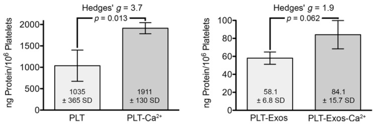
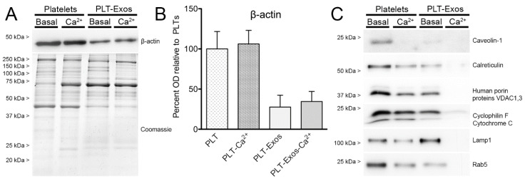
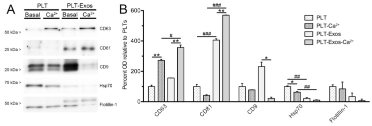
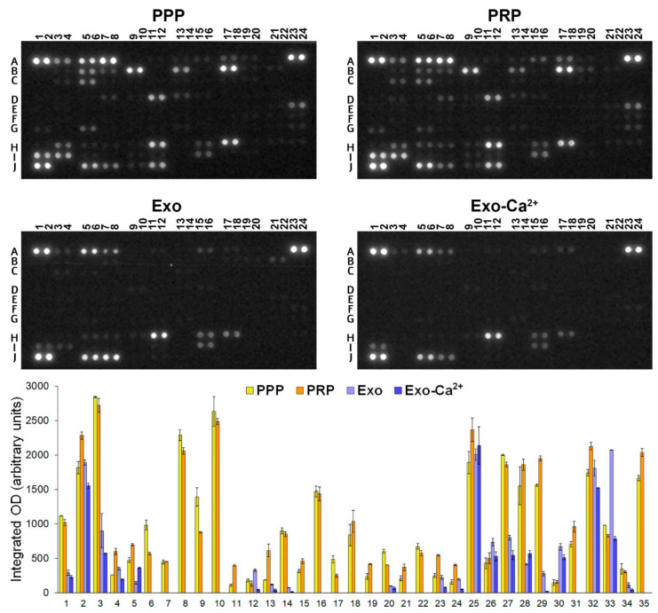
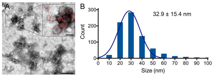
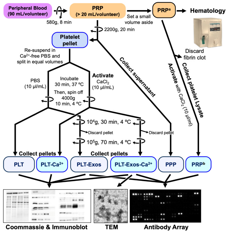
Platelet-Rich Plasma (PRP) is enriched in molecular messengers with restorative effects on altered tissue environments. Upon activation, platelets release a plethora of growth factors and cytokines, either in free form or encapsulated in exosomes, which have been proven to promote tissue repair and regeneration. Translational research on the potential of exosomes as a safe nanosystem for therapeutic cargo delivery requires standardizing exosome isolation methods along with their molecular and morphological characterization. With this aim, we isolated and characterized the exosomes released by human PRP platelets. Western blot analysis revealed that CaCl2-activated platelets (PLT-Exos-Ca2+) released more exosomes than non-activated ones (PLT-Exos). Moreover, PLT-Exos-Ca2+ exhibited a molecular signature that meets the most up-to-date biochemical criteria for platelet-derived exosomes and possessed morphological features typical of exosomes as assessed by transmission electron microscopy. Array analysis of 105 analytes including growth factors and cytokines showed that PLT-Exos-Ca2+ exhibited lower levels of most analytes compared to PLT-Exos, but relatively higher levels of those consistently validated as components of the protein cargo of platelet exosomes. In summary, the present study provides new insights into the molecular composition of human platelet-derived exosomes and validates a method for isolating highly pure platelet exosomes as a basis for future preclinical studies in regenerative medicine and drug delivery.

Keywords: cytokines; exosome markers; growth factors; human platelets; platelet-derived-exosomes; platelet-rich plasma.


Conflict of interest statement

The authors declare no conflict of interest. The funders had no role in the design of the study; in the collection, analyses, or interpretation of data; in the writing of the manuscript; or in the decision to publish the results.

Figures

Figure 1
Figure 1
Bar graphs representing the amount of protein, determined by the Bradford method and normalized to 106 platelets, in samples of platelets under basal (PLT) and calcium-stimulated (PLT-Ca2+) conditions and in samples of platelet-derived exosomes isolated from platelets at baseline (PLT-Exos) and after stimulation with calcium (PLT-Exos-Ca2+). Values shown correspond to mean values ± standard deviation. p values were calculated by Student’s unpaired (one-tail) t-test (PLT and PLT-Ca2+, n = 2; PLT-Exos and PLT-Exos-Ca2+, n = 3). Effect size was estimated by Hedges’ g.
Figure 2
Figure 2
Western blot analysis of non-exosome components in samples of human platelets and platelet-derived exosomes. (A) Protein loading controls by immunoblotting using an antibody against the cytoskeletal protein β-actin and by Coomassie Blue-staining of total proteins in platelet (PLT) samples, either non-activated (basal) or calcium-activated (Ca2+), and in platelet-derived exosomes (PLT-Exos) isolated from platelets at baseline or after stimulation with calcium. The immunoblot for β-actin corresponds to one of the two independent assays performed on samples from one volunteer. Coomassie blue staining was performed on samples pooled from volunteers 1 and 2. (B) Bar graph depicts the mean optical density values of immunoreactive bands for β-actin in the different samples relative to non-activated platelets. PLT, non-activated platelets; PLT-Ca2+, calcium-activated platelets; PLT-Exos, exosomes isolated from non-activated platelets; PLT-Exos-Ca2+, exosomes isolated from calcium-activated platelets. Data are mean ± SD of two independent experiments performed on samples from volunteers 1 and 2 (n = 2). (C) Immunoblot against proteins of non-exosome cell components (Table 2, for details) in PLT, PLT-Ca2+, PLT-Exos, and PLT-Exos-Ca2+ samples pooled from volunteers 1 and 2.
Figure 3
Figure 3
Biochemical analysis of the enrichment of platelet-derived exosome samples in exosome-specific markers. (A) Immunoblot against exosome-specific markers (see Table 2 for details) in platelet (PLT) samples, either non-activated (basal) or calcium-activated (Ca2+), and in platelet-derived exosomes (PLT-Exos) isolated from platelets at baseline or after stimulation with calcium. The immunoblot corresponds to one of the two independent assays performed on samples from one volunteer. (B) Bar graph depicts the mean optical density values of immunoreactive bands for five antigens widely used as exosome markers in the different samples relative to non-activated platelets. PLT, non-activated platelets; PLT-Ca2+, calcium-activated platelets; PLT-Exos, exosomes isolated from non-activated platelets; PLT-Exos-Ca2+, exosomes isolated from calcium-activated platelets. Data are mean ± SD of two independent experiments performed on samples from volunteers 1 and 2 (n = 2). Asterisks (*) and hashes (#) refer to comparisons between basal and calcium-activation conditions for each of the samples (PLT versus PLT-Ca2+, PLT-Exos versus PLT-Exos-Ca2+) and between exosomes and platelets of the same condition (PLT versus PLT-Exos and PLT-Ca2+ versus PLT-Exos-Ca2+). * p < 0.05, ** p < 0.01, # p < 0.05, ## p < 0.01, ### p < 0.005. p values were calculated by one-way repeated measures ANOVA with Bonferroni post hoc test.
Figure 4
Figure 4
Analysis of cytokines, chemokines, and growth factors in samples of platelet-poor plasma (PPP), platelet lysate from activated platelet-rich plasma (PRP), exosomes isolated from non-activated platelets (Exo), and exosomes isolated from CaCl2-activated platelets (Exo-Ca2+). The numbers and letters in antibody arrays at the top of the figure depict coordinates corresponding to the different analyte duplicates shown in Table 2. The bar graph at the bottom of the figure shows integrated optical density (OD) values (mean ± SD) measured in duplicate (two spots per analyte). The correspondence between the numbers in abscissa and analytes corresponding to the different coordinates are shown in Table 2.
Figure 5
Figure 5
(A) Transmission electron microscopy depicts a representative transmission electron microscopy (TEM) image of PLT-Exos-Ca2+. Scale bar, 200 nm; scale bar in inset, 50 nm. (B) The bar graph shows the distribution diagram of the exosomes and the fitted Gaussian fit of the diameter values of exosomes as measured in the TEM images. The value on the upper right side of the graph represents the mean diameter ± SD of a total of 775 exosomes.
Figure 6
Figure 6
Flow diagram of the isolation of the different samples used in the present study from whole human blood of young adults and the techniques used for their analysis. a It consists of the plasma fraction above the sedimented red blood cells, but not including the buffy coat. b This fraction, referred to as PRP throughout the text, corresponds to the platelet lysate collected from the activated PRP.

References

    1. Cecerska-Heryć E., Goszka M., Serwin N., Roszak M., Grygorcewicz B., Heryć R., Dołęgowska B. Applications of the regenerative capacity of platelets in modern medicine. Cytokine Growth Factor Rev. :2021. doi: 10.1016/j.cytogfr.2021.11.003. in press. - DOI - PubMed
    1. Le A.D.K., Enweze L., DeBaun M.R., Dragoo J.L. Platelet-Rich Plasma. Clin. Sports Med. 2019;38:17–44. doi: 10.1016/j.csm.2018.08.001. - DOI - PubMed
    1. Sánchez M., Anitua E., Delgado D., Sanchez P., Prado R., Orive G., Padilla S. Platelet-rich plasma, a source of autologous growth factors and biomimetic scaffold for peripheral nerve regeneration. Expert Opin. Biol. Ther. 2017;17:197–212. doi: 10.1080/14712598.2017.1259409. - DOI - PubMed
    1. Bohren Y., Timbolschi D.I., Muller A., Barrot M., Yalcin I., Salvat E. Platelet-rich plasma and cytokines in neuropathic pain: A narrative review and a clinical perspective. Eur. J. Pain. 2021;26:43–60. doi: 10.1002/ejp.1846. - DOI - PubMed
    1. Delgado D., Bilbao A.M., Beitia M., Garate A., Sánchez P., González-Burguera I., Isasti A., De Jesús M.L., Zuazo-Ibarra J., Montilla A., et al. Effects of platelet-rich plasma on cellular populations of the central nervous system: The influence of donor age. Int. J. Mol. Sci. 2021;22:1725. doi: 10.3390/ijms22041725. - DOI - PMC - PubMed