. 2022 Apr 8;376(6589):163-169.
doi: 10.1126/science.abn8933. Epub 2022 Mar 10.

Structure of a Janus kinase cytokine receptor complex reveals the basis for dimeric activation

Affiliations

Structure of a Janus kinase cytokine receptor complex reveals the basis for dimeric activation

Caleb R Glassman et al. Science. .

Abstract

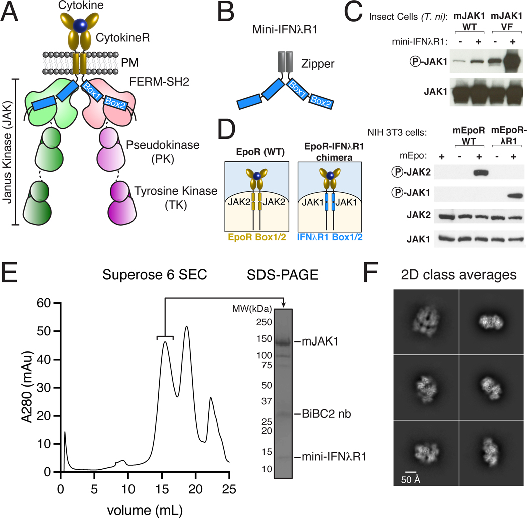
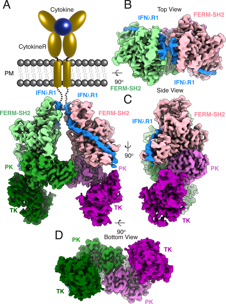
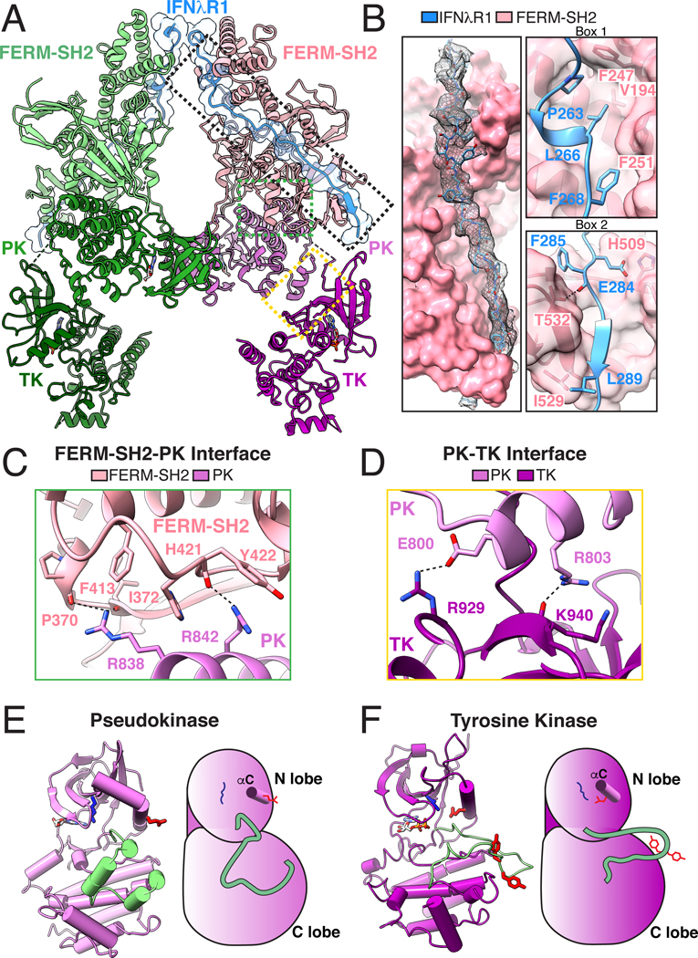
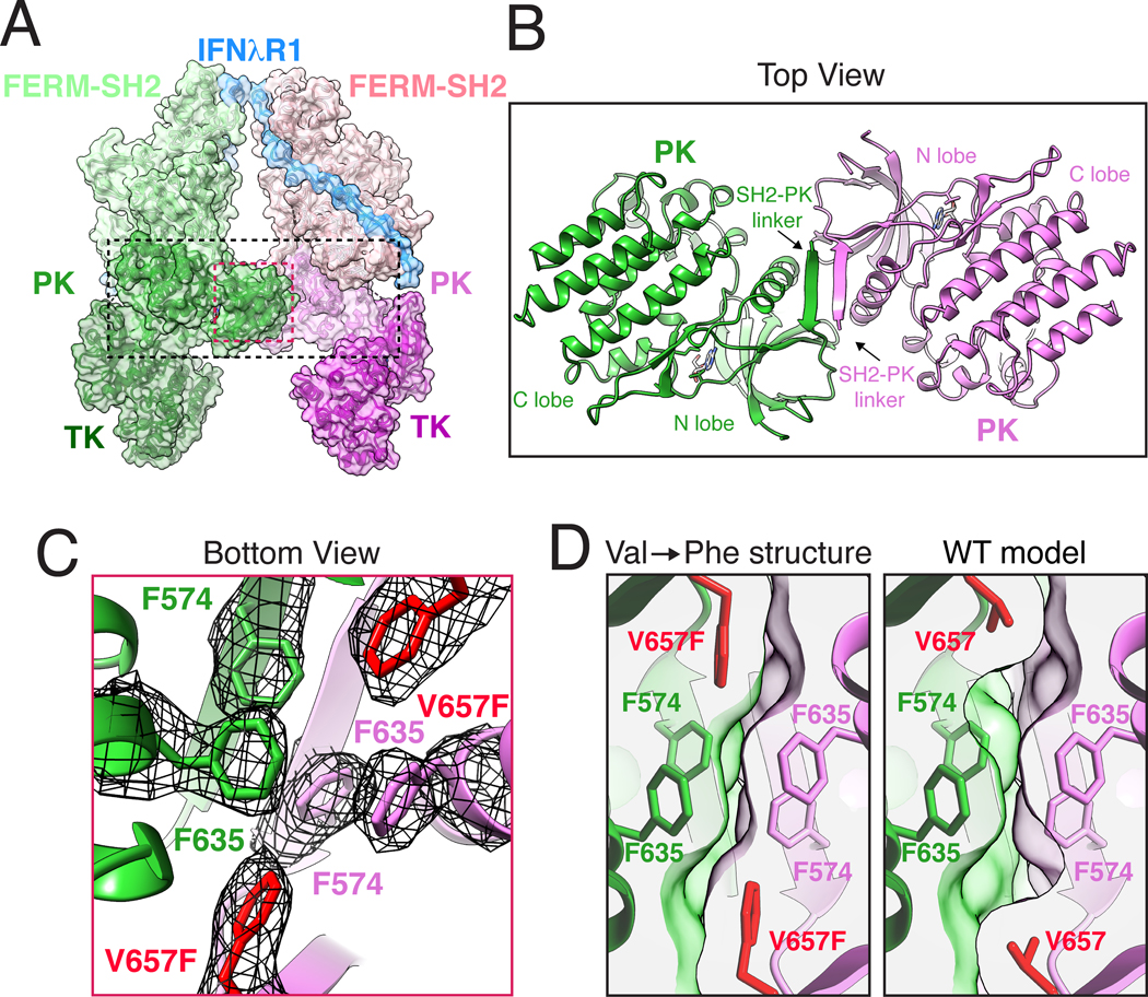
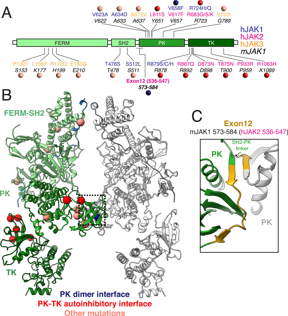
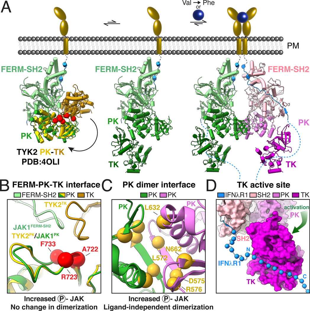
Cytokines signal through cell surface receptor dimers to initiate activation of intracellular Janus kinases (JAKs). We report the 3.6-angstrom-resolution cryo-electron microscopy structure of full-length JAK1 complexed with a cytokine receptor intracellular domain Box1 and Box2 regions captured as an activated homodimer bearing the valine→phenylalanine (VF) mutation prevalent in myeloproliferative neoplasms. The seven domains of JAK1 form an extended structural unit, the dimerization of which is mediated by close-packing of the pseudokinase (PK) domains from the monomeric subunits. The oncogenic VF mutation lies within the core of the JAK1 PK interdimer interface, enhancing packing complementarity to facilitate ligand-independent activation. The carboxy-terminal tyrosine kinase domains are poised for transactivation and to phosphorylate the receptor STAT (signal transducer and activator of transcription)-recruiting motifs projecting from the overhanging FERM (four-point-one, ezrin, radixin, moesin)-SH2 (Src homology 2)-domains. Mapping of constitutively active JAK mutants supports a two-step allosteric activation mechanism and reveals opportunities for selective therapeutic targeting of oncogenic JAK signaling.


Figures

Fig. 1.
Fig. 1.. Purification and biochemical characterization of an active JAK1-IFNλR1 complex.
(A) Schematic of a cell-surface, ligand-induced cytokine receptor-JAK dimer. (B) Schematic of a soluble cytokine receptor dimer mimetic (‘mini-IFNλR1’) consisting of N-terminal glutathione S-transferase (GST) tag for affinity purification, 3C protease site, GCN4-zipper, and mIFNλR1 Box1/Box2. (C) Mini-IFNλR1 expression enhances JAK1 phosphorylation when co-expressed in insect cells. Wild-type (WT) or Val657→Phe (VF) JAK1 was co-expressed with mini-IFNλR1 in T. ni cells by baculovirus transduction. JAK phosphorylation and total expression were measured two days post-infection by immunoblot of whole cell lysate. Results are representative of more than two independent experiments. (D) Schematic of wild-type (WT) EpoR/Epo complex (left), and EpoR-IFNλR1 chimera (right) showing substitution of Box1/Box2 motifs (blue). Cytokine-mediated dimerization of IFNλR1 Box1/Box2 results in JAK1 phosphorylation in mammalian cells. NIH 3T3 cells transiently expressing mEpoR or mEpoR-IFNλR1 chimera were stimulated with Epo for 20 min prior to analysis of JAK phosphorylation by immunoblot. Results are representative of two independent experiments. (E) Affinity-purification of JAK1 using zippered mini-IFNλR1 results in purification of a stable, non-aggregated complex. Superose 6 size exclusion chromatography (SEC, left) and sodium dodecyl sulphate–polyacrylamide gel electrophoresis (SDS-PAGE, right) of the JAK1-IFNλR1 complex. (F) Representative 2D class averages from single-particle cryo-EM imaging of the JAK1-IFNλR1 complex. PM – plasma membrane, R – receptor, mAu - milli-absorbance units, BiBC2 nb – tandem BC2 nanobody.
Fig. 2.
Fig. 2.. Cryo-EM structure of the active JAK1-IFNλR1 dimer.
(A) Segmented density map of the JAK1-IFNλR1 dimer resolved to 3.6Å resolution with extracellular and transmembrane domains shown as schematic. Subsequent panels show top (B), side (C), and bottom (D) views of the complex. Map threshold used in ChimeraX is set to 0.2 (~5.2σ). Individual JAK monomers are colored as a light-to-dark gradient from N to C terminus. Monomer 1: FERM-SH2, light green; PK, green; TK, dark green. Monomer 2: FERM-SH2, pink; PK, orchid; TK, purple. Density corresponding to IFNλR1 is colored blue.
Fig. 3.
Fig. 3.. Reconstitution of the full-length JAK1-IFNλR1 signaling complex.
(A) Ribbon diagram of the 2:2 JAK1-IFNλR1 complex. Dashed boxes indicate magnified views in the subsequent panels. (B) IFNλR1 binds JAK1 FERM and SH2 domains via N-terminal Box1 and C-terminal Box2 motifs within the receptor intracellular domain. Left: overall interaction between IFNλR1 and FERM-SH2 shown in surface representation with peptide density from the cryo-EM map shown as black mesh contoured at ~6.1σ. Upper right: IFNλR1 Box1 motif binds JAK1 FERM domain via a conserved PXXLXF motif. Lower right: IFNλR1 Box2 motif forms an anti-parallel β sheet with βG1 in the JAK1 SH2 domain. Hydrogen bonds and salt bridges are shown as black dashed lines. (C) Interface view of the FERM-SH2-PK domains. (D) Close-up view of the PK-TK interaction. (E) Ribbon diagram (left) and schematic (right) of the PK domain in standard view. Residues corresponding to the activation loop in a functional tyrosine kinase are shown in pale green. Active site Lys621 is shown in blue, catalytic Glu636 on αC helix is shown in red. (F) Ribbon diagram (left) and schematic (right) of the TK domain in standard view. The TK activation loop is colored pale green with tyrosine residues Tyr1033 and Tyr1034 colored red. The catalytic Glu924 (red) facing inwards towards Lys907 (blue) in the kinase active site. Amino acid abbreviations: F, Phe; V, Val; P, Pro; L, Leu; H, His; E, Glu; I, Ile; Y, Tyr; R, Arg; K, Lys; T, Thr.
Fig. 4.
Fig. 4.. JAK1 dimerization is mediated by the pseudokinase domain and enhanced the by oncogenic Val→Phe mutation.
(A) Ribbon diagram of the JAK1-IFNλR1 complex with semi-transparent surface. Dashed boxes indicate magnified views in the subsequent panels (B) Top view of the PK dimer at the center of the active JAK1 complex. The structure is shown as ribbon diagram with nucleotides shown as sticks. Labels indicate PK N lobe, C lobe, and SH2-PK linker. (C) Bottom view of the Phe triad with the cryo-EM density shown as black mesh contoured at ~9σ. Oncogenic V657F mutation is highlighted in red. (D) V657F enhances shape complementarity of the PK dimerization interface. Cross-section view of the PK-PK interface as seen from the bottom with V657F cryo-EM structure compared to a model of wild-type (WT) Val657. The WT model was created using Coot and surface clipping was set at Phe/Val657 Cβ for both panels to facilitate comparison. Amino acid abbreviations: F, Phe; V, Val.
Fig 5.
Fig 5.. Mapping human Gain of Function mutations on JAK1 suggests multiple mechanisms of oncogenic activation.
(A) Linear diagram of JAK domains showing the location of human gain of function mutations. Location of patient mutations in hJAK1 (blue), hJAK2 (pink), and hJAK3 (yellow) are shown above the analogous position in mJAK1 (44). Colored circles indicate classification of mutations based on their location at the active PK dimer interface (blue), in the autoinhibitory PK-TK interface based on a previously reported crystal packing structure of TYK2 (red) (18), or at sites with unknown function (salmon). (B) Structure of the active JAK1-IFNλR1 complex with the position of oncogenic mutations shown as balls colored by proposed mechanism of action as described above. (C) Close up of the PK dimer interface highlighting the residues in mJAK1 corresponding to hJAK2 exon 12 which has previously been identified as a hotspot for oncogenic mutations. Amino acid abbreviations: P, Pro; T, Thr; S, Ser; L, Leu; K, Lys; R, Arg; Q, Gln; E, Glu; G, Gly; C, Cys; D, Asp; N, Asn; V, Val; A, Ala; H, His; Y, Tyr; F, Phe.
Fig 6.
Fig 6.. Mechanistic model for JAK activation by both cytokine and oncogenic mutation.
(A) Proposed mechanism of JAK activation by ligand-induced dimerization and Val→Phe oncogenic mutation. An autoinhibited model of full-length JAK (left) was generated by docking a crystal structure of the PK-TK domains from hTYK2 (PDB ID: 4OLI; PK, yellow; TK, gold) (18) into the FERM-SH2-PK from the mJAK1 cryo-EM structure. Red balls indicate the position of activating mutations in the proposed autoinhibitory interface (44). A dynamic equilibrium between the autoinhibited ‘closed’ state and a partially active ‘open’ state (middle) exposes the PK domain and SH2-PK linker to allow for JAK dimerization. Cytokine-mediated receptor dimerization or oncogenic Val→Phe mutation facilitates formation of the PK dimer, sterically preventing autoinhibition and liberating the kinase domains for phosphotransferase activity (right). (B-C) Mechanistic mutations tracking receptor dimerization and JAK2 phosphorylation support a two-step model for activation. (B) Mutations at the proposed autoinhibitory interface enhance JAK2 phosphorylation but do not impact dimerization. Close up view of the autoinhibitory model in A with red balls indicate the positions of mutations previously found to increase JAK2 phosphorylation without inducing receptor dimerization (3). Residues are labeled according to their position in mJAK1: Ala722 (JAK2 Ile682→Phe), Arg723 (JAK2 Arg683→Gly), Phe733 (JAK2 Phe694→Leu). (C) Mutations at the PK dimerization interface increase both JAK2 phosphorylation and dimerization. Close up view of the JAK1-IFNλR1 PK dimer interface as viewed from the bottom. Yellow balls indicate the positions of mutations previously found to increase both JAK2 phosphorylation and receptor dimerization (3). Residues are numbered according to their position in mJAK1: Leu572 (JAK2 M535→Ile), Asp575 (JAK2 His538→Leu), Arg576 (JAK2 Lys539→Leu), Leu632 (JAK2 Glu592→Trp), Asn662 (JAK2 Asn622→Ile). (D) Model of receptor phosphorylation by the JAK1 dimer. Cryo-EM structure of the JAK1-IFNλR1 dimer is shown with TK domain in standard view. JAK1 is shown as surface with additional residues of IFNλR1 modeled as Cα balls for every other residue exiting the JAK1 SH2 domain and projecting towards the kinase active site. Amino acid abbreviations: F, Phe; A, Ala; R, Arg; L, Leu; N, Asn; D, Asp.

Comment in

References

    1. O’Shea JJ, Holland SM, Staudt LM, JAKs and STATs in immunity, immunodeficiency, and cancer. N Engl J Med 368, 161–170 (2013). - PMC - PubMed
    1. Wang X, Lupardus P, Laporte SL, Garcia KC, Structural biology of shared cytokine receptors. Annu Rev Immunol 27, 29–60 (2009). - PMC - PubMed
    1. Wilmes S. et al., Mechanism of homodimeric cytokine receptor activation and dysregulation by oncogenic mutations. Science 367, 643–652 (2020). - PMC - PubMed
    1. Stroud RM, Wells JA, Mechanistic diversity of cytokine receptor signaling across cell membranes. Sci STKE 2004, re7 (2004). - PubMed
    1. Watowich SS et al., Homodimerization and constitutive activation of the erythropoietin receptor. Proc Natl Acad Sci U S A 89, 2140–2144 (1992). - PMC - PubMed