. 2022 Mar 15;119(11):e2122161119.
doi: 10.1073/pnas.2122161119. Epub 2022 Mar 10.

The C terminus of the mycobacterium ESX-1 secretion system substrate ESAT-6 is required for phagosomal membrane damage and virulence

Affiliations

The C terminus of the mycobacterium ESX-1 secretion system substrate ESAT-6 is required for phagosomal membrane damage and virulence

Morwan M Osman et al. Proc Natl Acad Sci U S A. .

Abstract

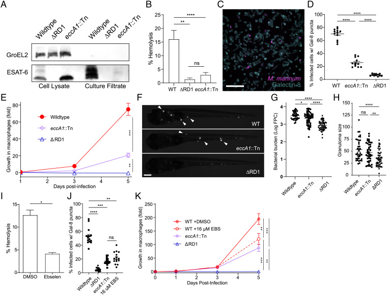
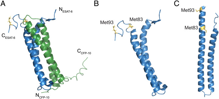
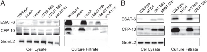
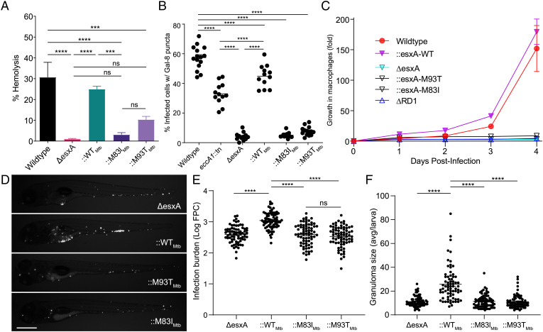
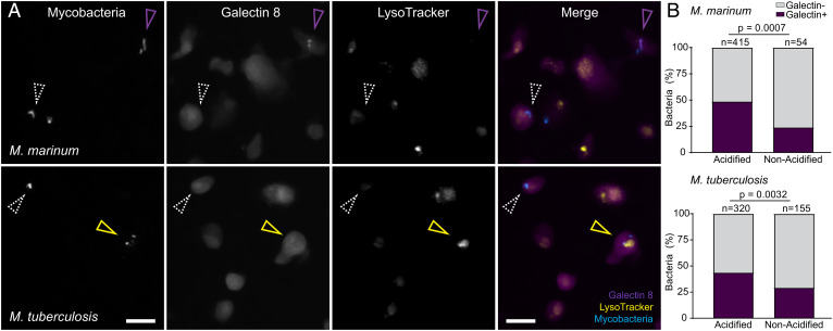
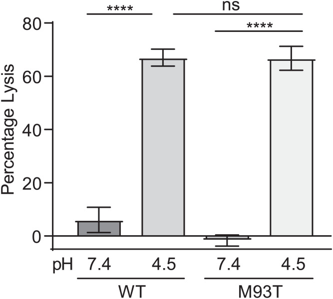
SignificanceTuberculosis (TB), an ancient disease of humanity, continues to be a major cause of worldwide death. The causative agent of TB, Mycobacterium tuberculosis, and its close pathogenic relative Mycobacterium marinum, initially infect, evade, and exploit macrophages, a major host defense against invading pathogens. Within macrophages, mycobacteria reside within host membrane-bound compartments called phagosomes. Mycobacterium-induced damage of the phagosomal membranes is integral to pathogenesis, and this activity has been attributed to the specialized mycobacterial secretion system ESX-1, and particularly to ESAT-6, its major secreted protein. Here, we show that the integrity of the unstructured ESAT-6 C terminus is required for macrophage phagosomal damage, granuloma formation, and virulence.

Keywords: ESAT-6; ESX-1; phagosomal damage; virulence.


Conflict of interest statement

Competing interest statement: R.B. is a coauthor on a 2018 paper with Eric Rubin, one of the reviewers of this paper.

Figures

Fig. 1.
Fig. 1.
Minimal ESX-1 secretion is required for intramacrophage survival and virulence. (A) Immunoblot of 48-h Mm cell lysates and culture filtrates. Data are representative of three independent experiments. GroEL2 is shown as a lysis control. (B) Contact-dependent hemolysis of RBCs by Mm. Data are combined from four experimental replicates. (C) Representative image of THP-1 cells infected with Mm, stained for galectin-8. (Scale bar, 100 μm.) (D) Percent of infected THP-1 macrophages with galectin-8 puncta. Each data point represents an individual imaging field. Horizontal lines, means. Statistics: One-way ANOVA with Šidák’s multiple comparisons test. (E) Intramacrophage growth of Mm within J774A.1 cells as measured by bacterial fluorescence. Data are representative of three independent experiments. (FH) Zebrafish larvae at 5 d postinfection. Data are representative of four independent experiments. (F) Representative images. (Scale bar, 250 μm.) Arrowheads, granulomas. (G) Bacterial burdens as assessed by bacterial fluorescence. (H) Average infection foci size per larva. Statistics: One-way ANOVA with Dunnett’s test. (I) Contact-dependent hemolysis of RBCs by Mm treated with vehicle or 16 μM ebselen. Data are combined from four experimental replicates. (J) Percent of infected THP-1 macrophages with galectin-8 puncta. Each data point represents an individual imaging field. Horizontal lines, means. Statistics: One-way ANOVA with Šidák’s multiple comparisons test. (K) Intramacrophage growth of J774A.1 cells infected with Mm. Data are representative of three independent experiments. (E and K) One-way ANOVA with Bonferroni’s multiple comparisons test. Statistics: ****P < 0.0001, ***P ≤ 0.001, **P < 0.01, *P < 0.05; not significant (ns), P > 0.05. WT, wild type.
Fig. 2.
Fig. 2.
ESAT-6 structures with C-terminal point mutations. (A) NMR structure of the heterodimer formed by ESAT-6 (blue) and CFP-10 (green) (Protein Data Bank [PDB] 1WA8). N and C termini of ESAT-6 and CFP-10 are as labeled, as well as methionine 83 and 93 of ESAT-6 (yellow). (B and C) Structure of ESAT-6 alone with methionine residues 83 and 93 highlighted, as determined experimentally by NMR (B) or by the predicted model using AlphaFold2 (C).
Fig. 3.
Fig. 3.
C-terminal point mutations in ESAT-6 support substantial levels of ESAT-6 and CFP-10 secretion. Immunoblots of 48-h cell lysates and culture filtrates of (A) Mm−ΔesxA and (B) Mm−ΔRD1 mutants. Each panel representative of three independent experiments. B is reprinted with permission from ref. .
Fig. 4.
Fig. 4.
ESAT-6 mediates phagosomal damage and virulence. (A) Contact-dependent hemolysis of RBCs by Mm. Data are combined from four experimental replicates. (B) Percent of infected THP-1 macrophages with galectin-8 puncta. Each data point represents individual imaging fields. Horizontal lines, means. Statistics: one-way ANOVA with Šidák correction for multiple comparisons. (C) Intramacrophage growth of Mm within J774A.1 macrophages as measured by bacterial fluorescence. Data are representative of four independent experiments. (D–F) Zebrafish larvae at 5 dpi. (D) Representative images. (Scale bar, 500 μm.) (E) Bacterial burdens as assessed by bacterial fluorescence. Statistics: One-way ANOVA with Dunnett’s test. (F) Average infection foci size per larva. Statistics: One-way ANOVA with Dunnett’s test. Data are representative of three independent experiments. Statistics: ****P < 0.0001, ***P ≤ 0.001; not significant (ns), P > 0.05.
Fig. 5.
Fig. 5.
Mm- and Mtb-mediated phagosomal membrane damage is enhanced in acidified phagosomes. (A and B) Galectin-8 labeled, LysoTracker Red-stained THP-1 cells at 24 h postinfection with EBFP2-expressing Mm (Top) or Mtb (Bottom). (A) Representative images of mycobacteria, Galectin-8, or LysoTracker, shown individually or as a merge of all three channels. (Scale bar, 10 μm.) Magenta arrowhead, bacteria proximal to galectin-8 puncta. Yellow arrowhead, acidified bacteria proximal to galectin-8 puncta. White arrowhead, bacteria that did not induce galectin-8 puncta. (B) Percent of bacteria located proximal to galectin-8 puncta, sorted by colocalization with LysoTracker positive compartments. Statistics: Fisher’s exact test.
Fig. 6.
Fig. 6.
Recombinant ESAT-6–M93T has wild-type levels of acidified liposome lysis activity. Quantification of pH-dependent liposome lysis by 3 µM recombinant ESAT-6 as measured by fluorescent ANTS release from DOPC liposomes. Statistics: One-way ANOVA with Šidák’s multiple comparisons test, ****P < 0.0001; not significant (ns), P > 0.05. Data are combined from four experimental replicates.

Similar articles

Cited by

References

    1. Lewis K. N., et al. , Deletion of RD1 from Mycobacterium tuberculosis mimics bacille Calmette-Guérin attenuation. J. Infect. Dis. 187, 117–123 (2003). - PMC - PubMed
    1. Pym A. S., Brodin P., Brosch R., Huerre M., Cole S. T., Loss of RD1 contributed to the attenuation of the live tuberculosis vaccines Mycobacterium bovis BCG and Mycobacterium microti. Mol. Microbiol. 46, 709–717 (2002). - PubMed
    1. Volkman H. E., et al. , Tuberculous granuloma formation is enhanced by a mycobacterium virulence determinant. PLoS Biol. 2, e367 (2004). - PMC - PubMed
    1. Brodin P., Rosenkrands I., Andersen P., Cole S. T., Brosch R., ESAT-6 proteins: Protective antigens and virulence factors? Trends Microbiol. 12, 500–508 (2004). - PubMed
    1. Mahairas G. G., Sabo P. J., Hickey M. J., Singh D. C., Stover C. K., Molecular analysis of genetic differences between Mycobacterium bovis BCG and virulent M. bovis. J. Bacteriol. 178, 1274–1282 (1996). - PMC - PubMed

MeSH terms

Substances