. 2022 May 2;132(9):e140685.
doi: 10.1172/JCI140685.

Macrophage-produced VEGFC is induced by efferocytosis to ameliorate cardiac injury and inflammation

Affiliations

Macrophage-produced VEGFC is induced by efferocytosis to ameliorate cardiac injury and inflammation

Kristofor E Glinton et al. J Clin Invest. .

Abstract

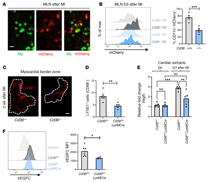
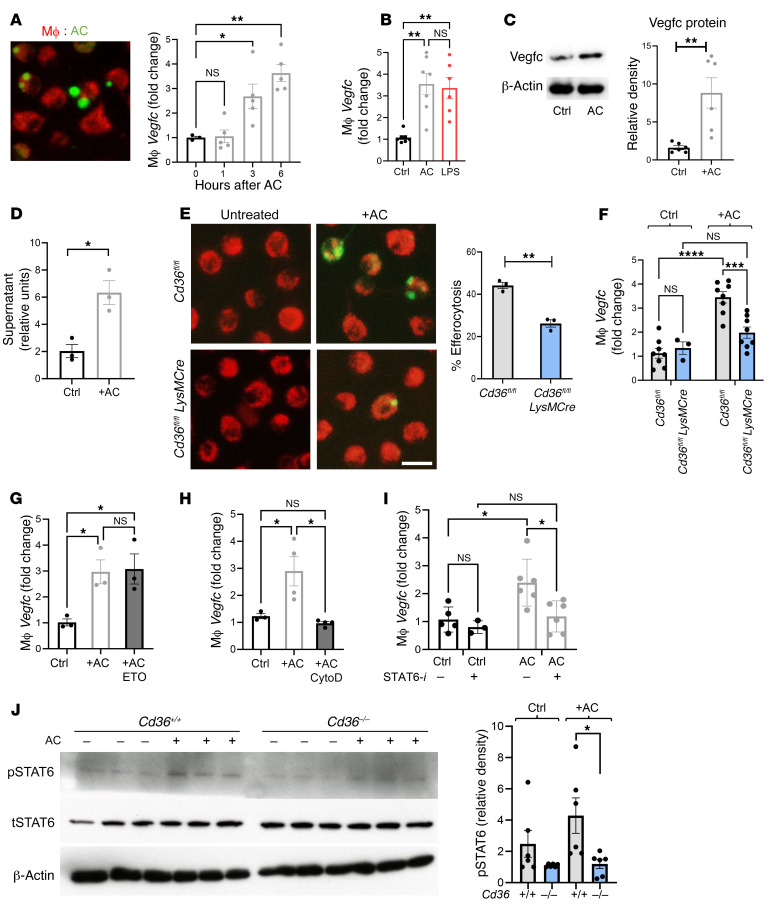
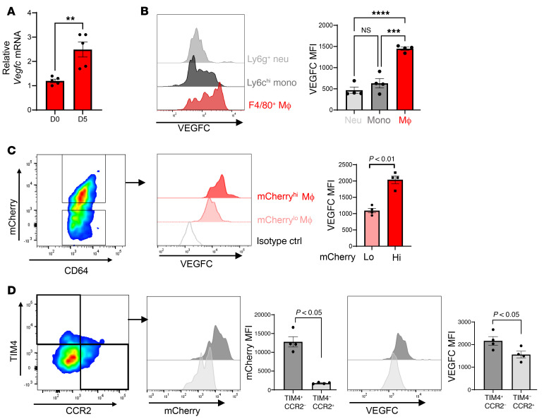
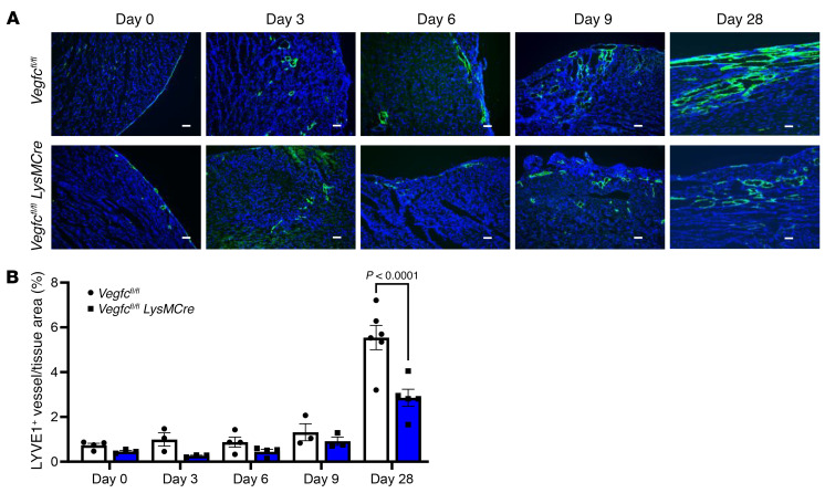
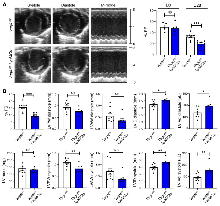
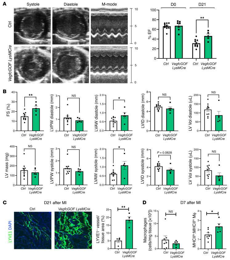
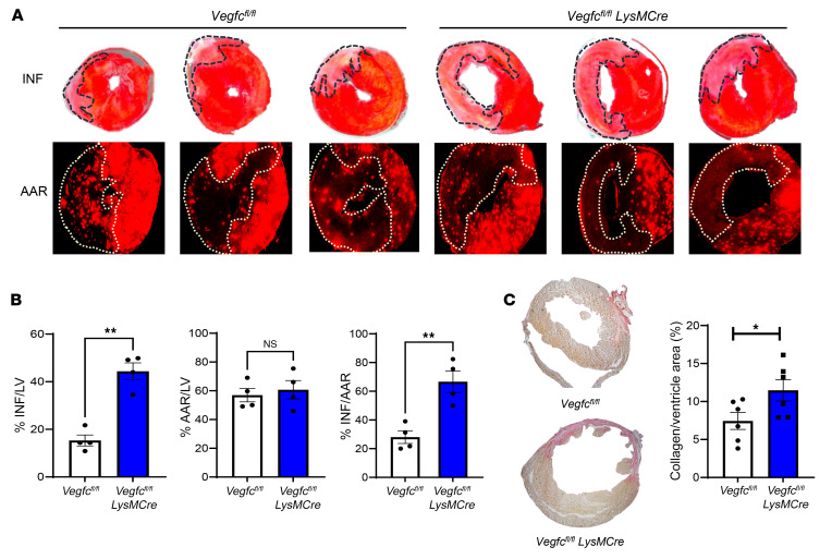
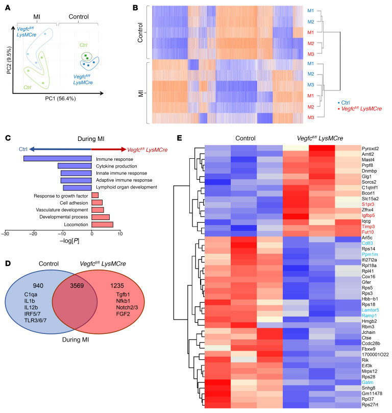
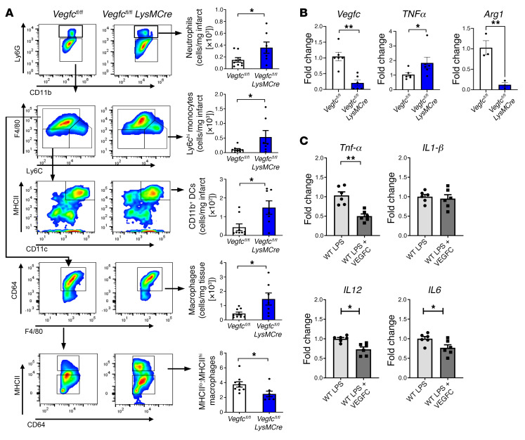
Clearance of dying cells by efferocytosis is necessary for cardiac repair after myocardial infarction (MI). Recent reports have suggested a protective role for vascular endothelial growth factor C (VEGFC) during acute cardiac lymphangiogenesis after MI. Here, we report that defective efferocytosis by macrophages after experimental MI led to a reduction in cardiac lymphangiogenesis and Vegfc expression. Cell-intrinsic evidence for efferocytic induction of Vegfc was revealed after adding apoptotic cells to cultured primary macrophages, which subsequently triggered Vegfc transcription and VEGFC secretion. Similarly, cardiac macrophages elevated Vegfc expression levels after MI, and mice deficient for myeloid Vegfc exhibited impaired ventricular contractility, adverse tissue remodeling, and reduced lymphangiogenesis. These results were observed in mouse models of permanent coronary occlusion and clinically relevant ischemia and reperfusion. Interestingly, myeloid Vegfc deficiency also led to increases in acute infarct size, prior to the amplitude of the acute cardiac lymphangiogenesis response. RNA-Seq and cardiac flow cytometry revealed that myeloid Vegfc deficiency was also characterized by a defective inflammatory response, and macrophage-produced VEGFC was directly effective at suppressing proinflammatory macrophage activation. Taken together, our findings indicate that cardiac macrophages promote healing through the promotion of myocardial lymphangiogenesis and the suppression of inflammatory cytokines.

Keywords: Inflammation; Innate immunity; Macrophages; Molecular pathology; Vascular Biology.


Figures

Figure 1
Figure 1. Bone marrow–derived Cd36 is required for both the accumulation of cardiac antigens in MLNs and increased tubular LYVE1 staining after MI.
(A) Imaging of murine MLN cross-sections 3 days after ligation of the LAD artery. Macrophages (MΦ) from LysMCre-EGFP mice show EGFP signal, and cardiomyocyte debris from Myh6-mCherry–transgenic mice show red signal. Scale bar: 40 μm. (B) Chimeric Myh6-mCherry mice, deficient for bone marrow–derived Cd36, were subjected to coronary artery occlusion. Three days after MI (D3), MLNs were harvested, and flow cytometric analysis of Ly6gCD11b+CD11c+ cells was performed. n = 5 per group. max, maximum. ***P < 0.0005, by 2-tailed, unpaired t test. (C) C57BL/6 mice were subjected to MI via coronary occlusion of the LAD artery. Representative immunofluorescence images were taken from 2 weeks after MI. Data display tubular LYVE1+ staining of the myocardial infarct border zone in Cd36–/– mice versus Cd36+/+ mice. Scale bar: 125 μm. (D) Border zone quantification of LYVE1+CD68 nuclei in Cd36fl/fl versus Cd36fl/fl LysMCre mice after MI. n = 5 per group. **P < 0.007, by 2-tailed, unpaired t test. (E) qPCR analysis of myocardial Vegfc in Cd36-deficient mice after MI. n = 5 per group. **P < 0.005 and ***P < 0.0001, by 2-way ANOVA followed by Tukey’s test. (F) Expression of VEGFC in cardiac macrophages with myeloid-specific deletion of Cd36 compared with controls 7 days after MI. n = 4–6 mice per group pooled from 2 independent experiments. *P < 0.05, by 2-tailed, unpaired t test.
Figure 2
Figure 2. Vegfc is induced in macrophages during efferocytosis.
(A) Photomicrograph depicts primary BMDMs (red) cocultivated with fluorescently labeled (green) ACs. Original magnification, ×40. Bar graph shows the quantification of gene expression at the indicated time points after AC cocultivation. n = 3–5. *P < 0.03 and **P < 0.003, by 1-way ANOVA with Tukey’s test. (B) Macrophage gene expression after treatment with either ACs or LPS (100 ng/mL). n = 6 per group. **P < 0.006. (C) Representative protein immunoblots of VEGFC, 6 hours after efferocytosis and densitometric analysis. n = 6 pooled from 2 independent experiments. **P < 0.005. (D) VEGFC ELISA of macrophage supernatant 9 hours after treatment with ACs versus control. n = 6. *P < 0.01. (E) BMDMs from Cd36+/+ and Cd36–/– animals were cocultured for 3 hours with ACs. After sequential washes, cells were imaged on an Olympus fluorescence microscope, and the percentage of efferocytosis was calculated from 10 random fields per replicate. Scale bar: 20 μm. Data are representative of 2 independent experiments with n = 3 wells per group. **P < 0.01. (F) BMDMs from Cd36+/+ and Cd3–/– animals was assessed for gene expression before and after treatment with ACs. n = 3–8 per group. ***P < 0.0005 and ****P < 0.0001. (G) Macrophage Vegfc gene expression after treatment with either ACs or with ACs plus etomoxir (ETO). n = 3–4 wells per group. *P < 0.01. (H) Quantification of gene expression in macrophages treated with ACs versus ACs plus cytochalasin D (CytoD). n = 3 per group. *P < 0.02. (I) Inhibition of STAT6 with AS1517499 blocked efferocytic Vegfc induction. *P < 0.01. (J) To assess STAT6 phosphorylation, macrophages were cultured as above, and lysates were prepared in RIPA buffer and then assessed by Western blotting. n = 6 pooled samples from 2 independent experiments. *P < 0.05. Data are presented as the mean ± SEM. Statistical significance was determined by 2-way ANOVA followed by Tukey’s test (B and FJ) and 2-tailed, unpaired t test (CE). Ctrl, control.
Figure 3
Figure 3. Selective expression of Vegfc in cardiac macrophages after MI.
(A) Sorted cardiac macrophages 5 days after MI were assessed for Vegfc mRNA expression compared with non-MI mice. n = 5/group. **P < 0.005, by unpaired t test. (B) Histogram of VEGFC expression in the indicated cell types by quantitative flow cytometry using cardiac extracts, 7 days after MI. Neutrophils (Neu) were defined as CD11b+Ly6g+; monocytes (Mono) were CD11b+Ly6gLy6chiF4/80; macrophages (Mac) were defined as CD11b+Ly6gLy6cloF4/80+CD64+. n = 4 per group. ***P < 0.0002 and ****P < 0.0001, by 2-way ANOVA followed by Tukey’s test. (C) mCherry mice were subjected to coronary ligation to track the uptake of cardiac antigens. Cardiac macrophages with higher levels of mCherry signal also expressed higher levels of VEGFC. n = 4 per group. P < 0.01, by 2-tailed, unpaired t test. (D) Cardiac macrophages were further classified by TIM4 (resident) or CCR2 (recruited) expression. TIM4+ resident macrophages had a higher frequency of mCherry uptake and expressed higher levels of VEGFC. n = 4 per group. Data were pooled from 2–3 independent experiments. P < 0.05, by 2-tailed, unpaired t test. Data are presented as the mean ± SEM.
Figure 4
Figure 4. Myeloid deletion of Vegfc reduces lymphatic vessel density after MI.
(A) Representative photomicrographs showing LYVE1 staining of coronary lymphatic vessels from Vegfcfl/fl LysMcre mice and Vegfcfl/fl littermates after permanent ligation of the LAD artery at the indicated time points. Scale bars: 50 μm. (B) Quantification of LYVE1 staining in myocardial ischemic AAR. P < 0.0001, by 2-way ANOVA followed by Tukey’s test. Data are presented as the mean ± SEM.
Figure 5
Figure 5. Myeloid Vegfc deficiency leads to impaired cardiac function after MI.
(A) Vegfcfl/fl LysMCre mice along with littermate Vegfcfl/fl controls were subjected to permanent ligation of the LAD artery. Representative M-mode still frames used for analysis showed a significant reduction in ventricular wall thickness and contraction in Vegfc-deficient animals. Parasternal short-axis M-mode measurements were collected prior to surgery (day 0) and again on day 28 after the ligation procedure. Using EF measurements as an indicator of cardiac function, no inherent differences were observed prior to injury, however, after 28 days, the Vegfc-deficient animals showed a significant reduction in EF. n = 9 control Vegfcfl/fl controls; n = 7 Vegfcfl/fl LysMCre mice. ***P < 0.005, by 2-tailed, unpaired t test. (B) Additional indices measured by echocardiography show significantly worsened indicators of systolic function including ventricular wall thickness, internal diameter, and volume. *P < 0.05, **P < 0.01, and ***P < 0.005, by 2-tailed, unpaired t test. FS, fractional shortening, LV Vol, left ventricular volume; LVID, left ventricular internal diameter; LVAW, left ventricular anterior wall thickness; LVPW, left ventricular posterior wall thickness.
Figure 6
Figure 6. Myeloid Vegfc overexpression leads to improved cardiac function and increased lymphangiogenesis after MI.
(A) VegfcGOF LysMCre mice and littermate controls were subjected to permanent ligation of the LAD artery. Parasternal short-axis M-mode measurements were collected prior to surgery (day 0) and again on day 21 after the ligation procedure to obtain EF measurements as an indicator of cardiac function. (B) Additional indices measured by echocardiography showed significantly improved indicators of systolic function including ventricular wall thickness, internal diameter, and volume. n = 9 control mice and n = 5 VegfcGOF LysMCre mice. (C) Representative photomicrographs and quantification of imaging from cardiac sections that were immunostained for LYVE1 in VegfcGOF LysMCre mice versus control, after MI. n = 4 per group. Original magnification, ×10. (D) Cardiac macrophages were assessed by flow cytometric analysis 7 days after MI. No significant differences in the absolute number of macrophages were observed. However, VegfcGOF LysMCre mice maintained a higher frequency of reparative MHCIIlo macrophages. *P < 0.05 and **P < 0.01, by 2-tailed, unpaired t test (AD).
Figure 7
Figure 7. Myeloid-derived Vegfc ameliorates scarring and infarct size after MI.
Mice of the indicated genotypes were subjected to experimental MI after ligation of the LAD artery. (A) The AAR was determined by intramyocardial circulation of fluorescent microbeads, and the infarct (INF) size was determined by TTC staining 7 days after the MI. (B) Quantification of the AAR and infarct size. n = 4 per group. **P < 0.0018, by 2-tailed, unpaired t test. (C) Representative Picrosirius red staining and quantification of fibrosis in cardiac sections on day 28 after the ligation procedure. n = 6 per group. *P < 0.05, by 2-tailed, unpaired t test.
Figure 8
Figure 8. Impaired immune response with myeloid Vegfc deficiency after MI.
Experimental C57BL/6 or Vegfcfl/fl versus Vegfcfl/fl LysMCre mice were subjected to coronary artery occlusion, and bulk mRNA gene expression analysis was performed for the LV. (A) Principal component analysis (PCA) revealing MI as a main source of variance in the data set. Data for the nonligated animals were clustered together, consistent with relatively comparable gene expression profiles at steady state. (B) Heatmap analysis and hierarchical clustering revealed distinct changes between nonligated and ligated animals at the transcriptional level. M1, M2, and M3 represent individual animals used for each group. (C) Gene ontology pathway interrogation revealed significant downregulation of immune response genes in the absence of myeloid Vegfc. In contrast, developmental pathways were induced in Vegfcfl/fl LysMCre mice, consistent with a hypertrophic response. (D) Venn diagram of differentially expressed or shared expression genes. (E) Heatmap of normalized top 50 absolute log fold changes in Vegfcfl/fl LysMCre mice compared with controls after MI. Genes highlighted in red are associated with inflammation and fibrosis, whereas those in cyan are associated with a lymphatic response and inflammation resolution.
Figure 9
Figure 9. Evidence for heightened cardiac and macrophage inflammation in myeloid Vegfc–deficient mice.
(A) Flow cytometric analysis of the ischemic AAR, 7 days after MI revealed heightened levels of CD11b+Ly6g+ neutrophils, Ly6chi monocytes, CD11b+CD11c+MHCIIhi DCs, and CD64+F4/80+ macrophages. Importantly, the ratio of MHCIIlo to MHCIIhi macrophages within the infarcted myocardium was significantly altered in Vegfc-deficient mice. n = 7–9 per group. *P < 0.05, by 2-tailed, unpaired t test. (B) BMDMs from Vegfcfl/fl and Vegfcfl/fl LysmCre mice were assessed for indicators of heightened inflammation. Transcript levels were measured by qPCR. An increase in markers of inflammation in Vegfc-deficient macrophages was observed along with reduced expression levels of Arg1. n = 3–6 per group. *P < 0.05 and **P < 0.004, by 2-tailed, unpaired t test. (C) VEGFC suppressed mRNA expression of inflammatory cytokines. qPCR of LPS-treated macrophages in culture treated with recombinant VEGFC versus control. n = 6 per group. *P < 0.05 and **P < 0.001, by 2-tailed, unpaired t test.

Comment in

Similar articles

Cited by

References

    1. Braunwald E. Biomarkers in heart failure. N Engl J Med. 2008;358(20):2148–2159. doi: 10.1056/NEJMra0800239. - DOI - PubMed
    1. Roger VL, et al. Trends in heart failure incidence and survival in a community-based population. JAMA. 2004;292(3):344–350. doi: 10.1001/jama.292.3.344. - DOI - PubMed
    1. McMurray JJ, et al. Which inhibitor of the renin-angiotensin system should be used in chronic heart failure and acute myocardial infarction? Circulation. 2004;110(20):3281–3288. doi: 10.1161/01.CIR.0000147274.83071.68. - DOI - PubMed
    1. Nahrendorf M, et al. Monocytes: protagonists of infarct inflammation and repair after myocardial infarction. Circulation. 2010;121(22):2437–2445. doi: 10.1161/CIRCULATIONAHA.109.916346. - DOI - PMC - PubMed
    1. van Amerongen MJ, et al. Macrophage depletion impairs wound healing and increases left ventricular remodeling after myocardial injury in mice. Am J Pathol. 2007;170(3):818–829. doi: 10.2353/ajpath.2007.060547. - DOI - PMC - PubMed

Publication types

Substances