. 2022 Apr 28;139(17):2601-2621.
doi: 10.1182/blood.2021014343.

International expert consensus recommendations for the diagnosis and treatment of Langerhans cell histiocytosis in adults

Affiliations

International expert consensus recommendations for the diagnosis and treatment of Langerhans cell histiocytosis in adults

Gaurav Goyal et al. Blood. .

Abstract

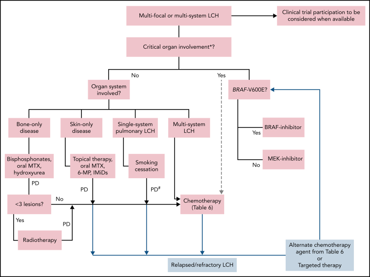
Langerhans cell histiocytosis (LCH) can affect children and adults with a wide variety of clinical manifestations, including unifocal, single-system multifocal, single-system pulmonary (smoking-associated), or multisystem disease. The existing paradigms in the management of LCH in adults are mostly derived from the pediatric literature. Over the last decade, the discovery of clonality and MAPK-ERK pathway mutations in most cases led to the recognition of LCH as a hematopoietic neoplasm, opening the doors for treatment with targeted therapies. These advances have necessitated an update of the existing recommendations for the diagnosis and treatment of LCH in adults. This document presents consensus recommendations that resulted from the discussions at the annual Histiocyte Society meeting in 2019, encompassing clinical features, classification, diagnostic criteria, treatment algorithm, and response assessment for adults with LCH. The recommendations favor the use of 18F-Fluorodeoxyglucose positron emission tomography-based imaging for staging and response assessment in the majority of cases. Most adults with unifocal disease may be cured by local therapies, while the first-line treatment for single-system pulmonary LCH remains smoking cessation. Among patients not amenable or unresponsive to these treatments and/or have multifocal and multisystem disease, systemic treatments are recommended. Preferred systemic treatments in adults with LCH include cladribine or cytarabine, with the emerging role of targeted (BRAF and MEK inhibitor) therapies. Despite documented responses to treatments, many patients struggle with a high symptom burden from pain, fatigue, and mood disorders that should be acknowledged and managed appropriately.


Figures

Figure 1
Figure 1
Spectrum of manifestations of LCH in adults. (A) Axial computed tomography (CT) of the calvarium demonstrating a lytic lesion with a beveled edge (arrow). (B) Sagittal pituitary T2 weighted fluid-attenuated inversion recovery (FLAIR) magnetic resonance imaging (MRI) highlighting a hypothalamic lesion with increased FLAIR signal (arrow). (C) Sagittal T1 weighted contrast-enhanced brain MRI demonstrating an enhancing posterior midbrain/pons lesion (circle). (D) Axial brain MRI with a neurodegenerative pattern of T2 signal abnormality throughout the bilateral cerebellar peduncles (brackets). (E-G) Axial CT of the mid to upper chest demonstrating variable appearances of pulmonary LCH to include the most common nodulocystic pattern ground-glass nodules and cysts (E), a cystic-predominant pattern with larger irregular cysts and a few scattered nodules (F), and more confluent combined cystic and nodular disease with architectural distortion consistent with elements of fibrosis (G). (H) Axial fused 18F-fluorodeoxyglucose positron emission tomography CT (FDG PET/CT) of the chest demonstrating 2 FDG avid left axillary lymph nodes (arrows). (I) Diffusion-weighted MRI of the liver demonstrating an infiltrative pattern of increased signal throughout the right hepatic lobe (brackets). (J) Magnetic resonance cholangiopancreatography demonstrating multifocal stricturing (arrow) and dilatation (bracket) of the intrahepatic ducts. (K) Maximum intensity projection FDG PET/CT demonstrating FDG avid advanced pulmonary disease (L), multifocal FDG avid hepatic disease (M), and an FDG avid lytic right femur lesion (N). (O) MIP FDG PET/CT demonstrating an infiltrative FDG avid laryngeal mass (P), bilateral axillary dermal lesions with associated left axillary photograph (Q), and a mildly FDG avid mesenteric mass highlighted with a bracket (R).
Figure 2
Figure 2
Histopathologic and immunophenotypic features of LCH. The characteristic infiltrate of LCH on hematoxylin and eosin shows mononuclear cells with grooved nuclei and abundant eosinophilic cytoplasm, with a mixture of eosinophils and small lymphocytes. The LCH cells show expression of CD68 (cytoplasmic, Golgi dot-like staining with background macrophages darkly stained), CD1a (surface), and langerin (cytoplasmic) and are negative for CD163. The mutant-specific antibody clone VE1 detects the BRAF-V600E mutation by immunohistochemistry with 2-3+ strong cytoplasmic staining. Images magnification ×400; inset magnification ×600.
Figure 3
Figure 3
Suggested workup for a newly diagnosed or suspected LCH in adults. ACTH, adrenocorticotropic hormone; CBC, complete blood count; DI, diabetes insipidus; EKG, electrocardiogram; FSH/LH, follicle stimulating hormone/luteinizing hormone; HEENT, head, eyes, ears, nose, and throat; IGF-1, insulin-like growth factor 1; TSH, thyroid-stimulating hormone.
Figure 4
Figure 4
Treatment algorithm for adults with multifocal or multisystem LCH. Systemic therapy is indicated for patients with single-system unifocal disease involving critical organs or specific sites (nervous system, liver, spleen, etc). *Brain (esp. neurodegenerative LCH), liver (esp. sclerosing cholangitis). Liver transplant consult for sclerosing cholangitis. #Systemic therapy may be indicated in patients with symptomatic disease and unable to quit smoking. Lung transplantation referral should be undertaken if not eligible for or refractory to systemic treatments. 6-MP, 6-mercaptopurine; IMiDs, immunomodulators (thalidomide, lenalidomide); MTX, methotrexate; PD, progressive disease.

References

    1. Swerdlow SH, Campo E, Harris NL, et al. WHO Classification of Tumours of Haematopoietic and Lymphoid Tissues. International Agency for Research on Cancer; Lyon, France: 2017.
    1. Badalian-Very G, Vergilio JA, Degar BA, et al. Recurrent BRAF mutations in Langerhans cell histiocytosis. Blood. 2010;116(11):1919–1923. - PMC - PubMed
    1. Emile JF, Abla O, Fraitag S, et al. Histiocyte Society Revised classification of histiocytoses and neoplasms of the macrophage-dendritic cell lineages. Blood. 2016;127(22):2672–2681. - PMC - PubMed
    1. Goyal G, Shah MV, Hook CC, et al. Adult disseminated Langerhans cell histiocytosis: incidence, racial disparities and long-term outcomes. Br J Haematol. 2018;182(4):579–581. - PubMed
    1. Makras P, Stathi D, Yavropoulou M, Tsoli M, Kaltsas G. The annual incidence of Langerhans cell histiocytosis among adults living in Greece. Pediatr Blood Cancer. 2020;67(9):e28422. - PubMed

Publication types