. 2022 Mar 11;13(1):5.
doi: 10.1186/s43008-022-00090-6.

A severe microsporidian disease in cultured Atlantic Bluefin Tuna (Thunnus thynnus)

Affiliations

A severe microsporidian disease in cultured Atlantic Bluefin Tuna (Thunnus thynnus)

Alejandro López-Verdejo et al. IMA Fungus. .

Abstract

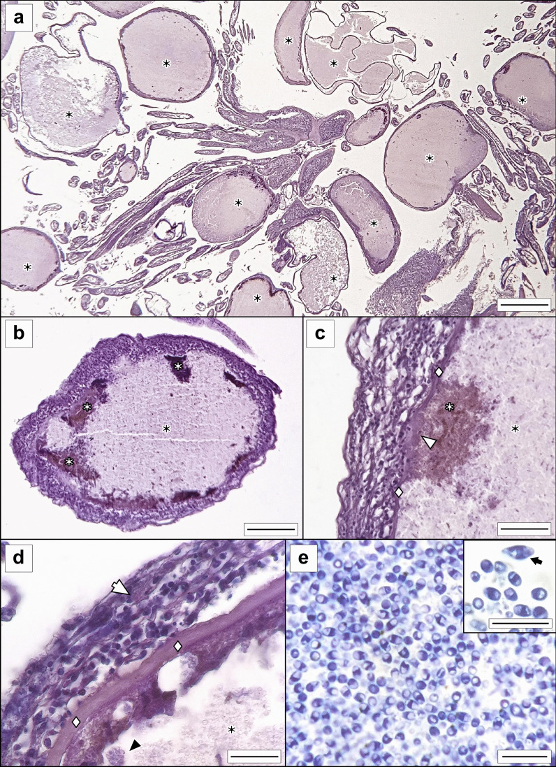
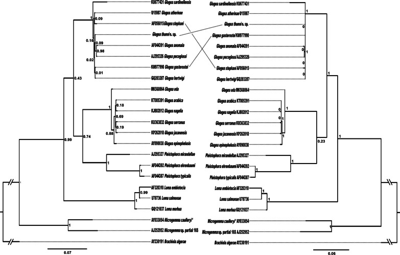
One of the most promising aquaculture species is the Atlantic bluefin tuna (Thunnus thynnus) with high market value; disease control is crucial to prevent and reduce mortality and monetary losses. Microsporidia (Fungi) are a potential source of damage to bluefin tuna aquaculture. A new microsporidian species is described from farmed bluefin tunas from the Spanish Mediterranean. This new pathogen is described in a juvenile associated with a highly severe pathology of the visceral cavity. Whitish xenomas from this microsporidian species were mostly located at the caecal mass and ranged from 0.2 to 7.5 mm. Light and transmission electron microscopy of the spores revealed mature spores with an average size of 2.2 × 3.9 μm in size and a polar filament with 13-14 coils arranged in one single layer. Phylogenetic analysis clustered this species with the Glugea spp. clade. The morphological characteristics and molecular comparison confirm that this is a novel microsporidian species, Glugea thunni. The direct life-cycle and the severe pathologies observed makes this parasite a hard risk for bluefin tuna cultures.

Keywords: Aquaculture; Glugea thunni new taxa; Glugeida; Marinosporidia; Microsporidia; Osteichthyes; Scombridae; Xenoma.


Conflict of interest statement

The authors declare that they have no competing interests.

Figures

Fig. 1
Fig. 1
Glugea thunni in Thunnus thynnus from the Mediterranean Sea. a Specimen of T. thynnus infected by G. thunni with melanized and partially melanized cysts, including a detail of xenomas within the ceacal mass (scale bar 2.5 cm) b fresh smear with free microspores and parasitophorous vesicles with different number of microspores (scale bar 40 µm). c Detail of fresh smear with free short and large microspores and parasitophorous vesicles (scale bar 20 µm). White arrow—melanized cyst; white arrowhead—cysts with melanized spots; black arrows—abnormal microspores; black arrowheads—parasitophorous vesicles
Fig. 2
Fig. 2
Micrographs of Glugea thunni from histological sections of the caecal mass of Thunnus thynnus from the Mediterranean Sea. a G. thunni xenomas in the mesentery among the intestinal caeca (scale bar 1 mm). b Cyst of G. thunni; xenoma exhibits peripheral spots with different degrees of melanization (scale bar 200 µm). c Detail of peripheral xenoma melanization (scale bar 70 µm). d Cyst wall with eosinophilic granule cell in the outer celular layer (scale bar 40 µm). e Microspores at the central region of xenoma (scale bar 10 µm) with a detail including an abnormal microspore (scale bar 5 µm) (ad, paraffin sections stained in H–W; e, semi-thin stained in toluidine blue). White arrow—eosinophilic granule cell; black asterisk—xenoma; white asterisk—melanized spot; white diamond—acellular/fibrous layer; white arrowhead—disintegrated acellular/fibrous layer; black arrowhead—parasitophorus vesicle
Fig. 3
Fig. 3
Transmission electron micrographs of xenomas of Glugea thunni from the caecal mass of Thunnus thynnus from the Mediterranean Sea. a Mature spores and degenerative host cells. b and c Spores under development. MS—mature spores; IS—immature spores; Sb—Early sporoblast; HN, Host cell nucleus; black arrowhead—parasitophorous vacuole membrane (scale bars 2 µm)
Fig. 4
Fig. 4
Transmission electron micrographs of Glugea thunni from the caecal mass of Thunnus thynnus from the Mediterranean Sea. a Longitudinal section of an adult microspore showing the ultrastructure (scale bar 500 nm). b Detail of the anchoring disk and spore wall (scale bar 400 nm). c Detail of polar filament surrounding the spore (scale bar 1 µm). Abbreviations: AD—anchoring disk; En—wall endospore; Ex—wall exospore; N—spore nucleus; PF—polar filament; Pp—polaroplast; PV—posterior vacuole; Wa—spore wall
Fig. 5
Fig. 5
Confronted bayesian inference (BI) and maximum likelihood (ML) trees for the analyses of the microsporidians based on partial 16S rDNA sequences (774 bp). Nodal support is given as posterior probabilities (BI) and bootstrap values resulting from maximum likelihood (ML); only values > 0.95 (BI) and 70% (ML) are shown. The scale-bars indicate the expected number of substitutions per site. *Microgemma caulleryi is accepted as G. microspora in Lom
Fig. 6
Fig. 6
Resulting tree for the analyses of the microsporidians based on partial 16S rDNA sequences (1713 bp). Nodal support is given as posterior probabilities (BI) and bootstrap values resulting from maximum likelihood (ML) analysis in the form (BI/ML); only values > 0.95 (BI) and 70% (ML) are shown. The scale-bar indicates the expected number of substitutions per site

References

    1. Abdel-Baki AAS, Al-Quraishy S, Rocha S, Dkhil MA, Casal G, Azevedo C. Ultrastructure and phylogeny of Glugea nagelia n. sp. (Microsporidia: Glugeidae), infecting the intestinal wall of the yellowfin hind, Cephalopholis hemistiktos (Actinopterygii: Serranidae), from the Red Sea. Folia Parasitologica. 2015;62(1):1–7. - PubMed
    1. Abdel-Baki AAS, Tamihi AF, Al-Qahtani HA, Al-Quraishy S, Mansour L. Glugea jazanensis sp. Nov. infecting Lutjanus bohar in the Red Sea: ultrastructure and phylogeny. Dis Aquat Organ. 2015;116(3):185–190. - PubMed
    1. APROMAR (2020) Aquaculture in Spain 2020 Asociación Empresarial de Productores de Cultivos Marinos. Chiclana (Cádiz). http://www.apromar.es/content/informes-anuales
    1. Azevedo C, Abdel-Baki AAS, Rocha S, Al-Quraishy S, Casal G. Ultrastructure and phylogeny of Glugea arabica n. sp. (Microsporidia), infecting the marine fish Epinephelus polyphekadion from the Red Sea. Eur J Protistol. 2016;52(228):11–21. - PubMed
    1. Bell AS, Aoki T, Yokoyama H. Phylogenetic relationships among microsporidia based on rDNA sequence data, with particular reference to fish-infecting Microsporidium Balbiani 1884 species. J Eukaryot Microbiol. 2001;48(3):258–265. - PubMed

LinkOut - more resources