Review
. 2022 May;43(5):362-377.
doi: 10.1016/j.tips.2022.02.007.

The current toolbox for APOBEC drug discovery

Affiliations
Review

The current toolbox for APOBEC drug discovery

Michael J Grillo et al. Trends Pharmacol Sci. 2022 May.

Abstract

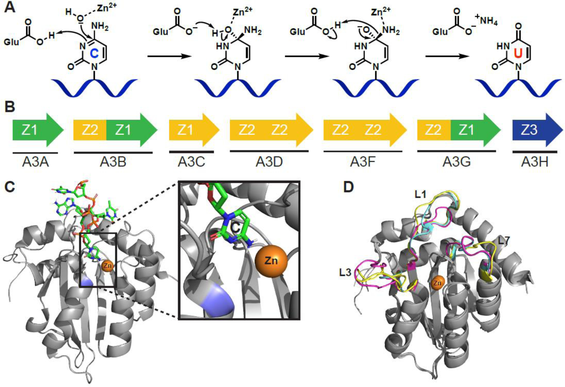
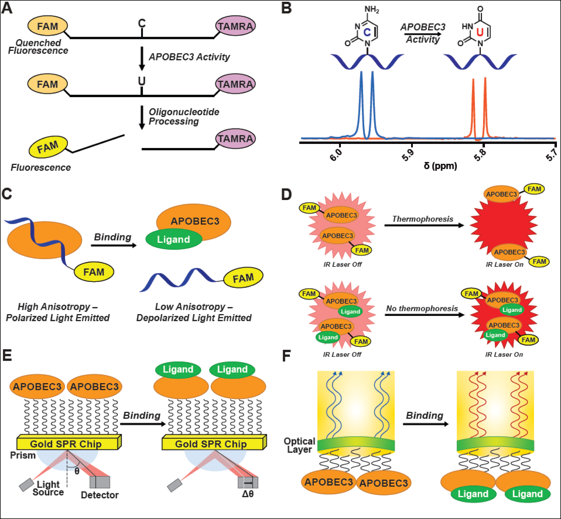
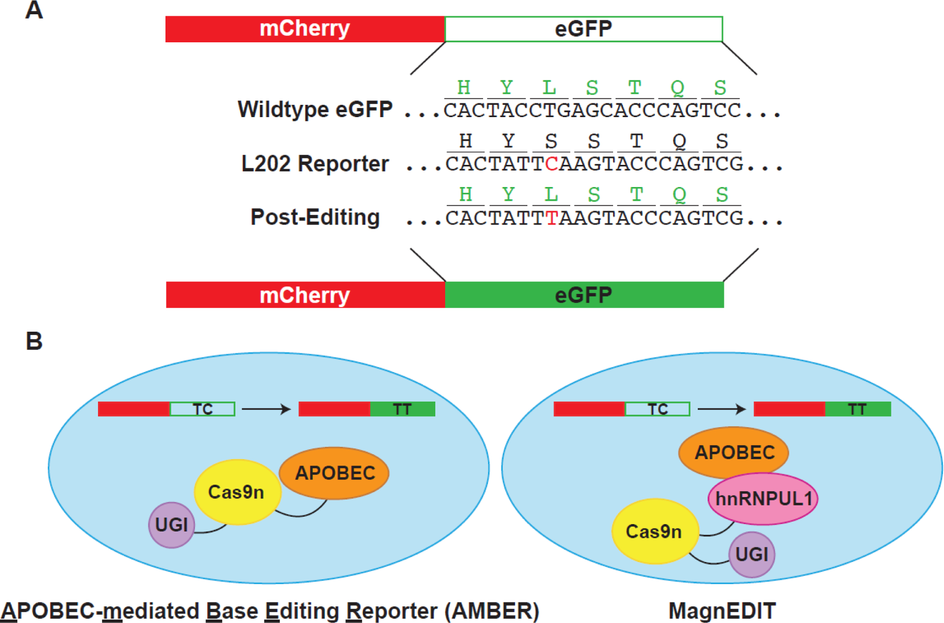
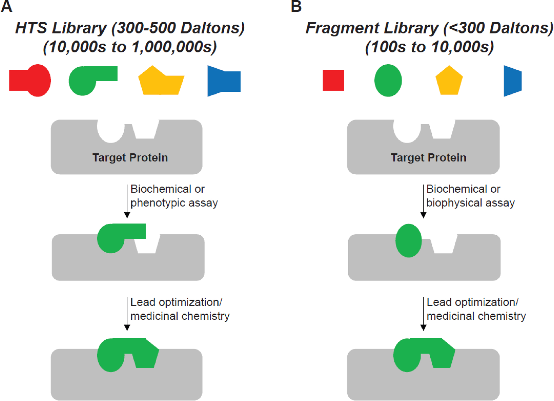
Mutational processes driving genome evolution and heterogeneity contribute to immune evasion and therapy resistance in viral infections and cancer. APOBEC3 (A3) enzymes promote such mutations by catalyzing the deamination of cytosines to uracils in single-stranded DNA. Chemical inhibition of A3 enzymes may yield an antimutation therapeutic strategy to improve the durability of current drug therapies that are prone to resistance mutations. A3 small-molecule drug discovery efforts to date have been restricted to a single high-throughput biochemical activity assay; however, the arsenal of discovery assays has significantly expanded in recent years. The assays used to study A3 enzymes are reviewed here with an eye towards their potential for small-molecule discovery efforts.

Keywords: APOBEC; DNA deaminase inhibitors; chemical probes; drug discovery; screening.


Conflict of interest statement

Declaration of interests R.S.H. and D.A.H. were cofounders of ApoGen Biotechnologies, Inc., which closed operations in April 2021. The other authors declare no conflicts of interest.

Figures

Figure 1.
Figure 1.
APOBEC3 structure and function. A) Proposed C-to-U hydrolytic deamination mechanism. B) The human APOBEC3 subfamily represented by arrows (single or double domain), and colors indicating phylogenetic grouping (single or double domain). C) A3B C-terminal domain co-crystal structure bound to ssDNA (PDB: 5TD5). The target cytosine is shown (inset) to be in proximity to the catalytic zinc (orange) and catalytic Glu255 (blue; mutated to Ala for crystallographic studies). D) Overlay of A3A (magenta, PDB: 4XXO), A3B C-terminal domain (cyan, PDB: 5CQI), and A3G C-terminal domain (yellow, PDB: 3IR2) showing conservationed structure between Z1 domains of different APOBEC3 enzymes of therapeutic relevance. Most of the structural variability occurs in structure is in the flexible loop regions that have significant effects on substrate binding and catalytic activity. Structure alignments performed with Pymol 2.3 align function.
Figure 2.
Figure 2.
Assays used for APOBEC3 drug discovery. A) Microplate-based APOBEC3 deamination activity assay. A ssDNA containing a target cytosine is labeled on the 5′ and 3′ end, respectively, with FAM and TAMRA (FAM emission quenched by FRET with TAMRA). When incubated with active APOBEC3 enzyme, the target cytosine is converted to a uracil, and treatment with UDG, then alkaline conditions yields a cleaved oligonucleotide with an increase in measurable FAM fluorescence. B) NMR-based APOBEC3 activity assay. The C5-proton signal is monitored by 1H-NMR. Upon deamination of the substrate, the signal is shifted, and deamination is quantified. C) Fluorescence anisotropy/polarization assay (FA/FP). Fluorescence is emitted by FAM on a labeled ssDNA that is subjected to polarized light. When bound to APOBEC3, the ssDNA tumbles slowly leaving the polarized light intact (high signal). When ssDNA is displaced, the faster tumbling depolarizes the light (low signal). D) Microscale thermophoresis (MST). FAM-labeled APOBEC3 moves through a capillary in an IR-beam-generated temperature gradient (thermophoresis). Binding of a ligand changes the APOBEC3 thermophoretic property, thereby preventing APOBEC3 from moving away from the irradiated region in the capillary, thus resulting in an increase in fluorescent signal when compared to the unbound state. E) Surface plasmon resonance (SPR). APOBEC3 is immobilized to a gold chip and ligand binding to the protein results in a quantifiable change in the angle of the reflected light source (θ). F) Biolayer interferometry (BLI). A dip-and-read sensor is functionalized with APOBEC3 and the interference pattern of incident white light through the sensor is measured following binding of a ligand to the APOBEC3 enzyme.
Figure 3.
Figure 3.
Overview of cellular assays for in vivo activity assessment. A) Plasmid editing by APOBEC-mediated Base Editing Reporter (AMBER). Reporter plasmid contains constitutively-expressed active mCherry for transfection/transduction normalization. eGFP reporter protein is inactivated by mutation of the L202 codon to encode for a serine residue, which ablates fluorescence. Silent mutations were also introduced near the editing site to reduce the opportunity for double-stranded breaks. APOBEC3 editing restores eGFP fluorescence. B) Comparison of AMBER editing system and MagnEDIT. AMBER fuses all three proteins – APOBEC3, Cas9n, and UGI – to direct editing to the eGFP reporter construct in panel A. MagnEDIT fuses Cas9n/UGI with an APOBEC-interacting protein, hnRNPUL1. gRNA directs hnRNPUL1/Cas9n/UGI to the eGFP reporter construct from panel A. hnRNPUL1 therefore acts as a “magnet” to attract the APOBEC3 protein to the reporter and restore eGFP fluorescence. Elimination of the tethering of the APOBEC3 protein to the Cas9n/UGI construct results in less off-target editing.
Figure I.
Figure I.
HTS and FBDD approaches. Depiction of typical library design parameters and types of assays used for A) HTS, and B) FBDD.

References

    1. Betts L, et al. (1994) Cytidine deaminase. The 2.3 Å crystal structure of an enzyme: transition-state analog complex. J. Mol. Biol. 235, 635–656 - PubMed
    1. Johansson E, et al. (2002) Crystal structure of the tetrameric cytidine deaminase from Bacillus subtilis at 2.0 Å resolution. Biochemistry 41, 2563–2570 - PubMed
    1. Yao L, et al. (2005) A molecular dynamics exploration of the catalytic mechanism of yeast cytosine deaminase. J. Phys. Chem. B 109, 7500–7510 - PubMed
    1. Hall RS, et al. (2011) Three-dimensional structure and catalytic mechanism of cytosine deaminase. Biochemistry 50, 5077–5085 - PMC - PubMed
    1. Manta B, et al. (2014) Reaction Mechanism of Zinc-Dependent Cytosine Deaminase from Escherichia coli: A Quantum-Chemical Study. J. Phys. Chem. B 118, 5644–5652 - PubMed

Publication types

Substances