Review
. 2022 Feb 22:13:828780.
doi: 10.3389/fendo.2022.828780. eCollection 2022.

Metabolic and Epigenetic Regulation by Estrogen in Adipocytes

Affiliations
Review

Metabolic and Epigenetic Regulation by Estrogen in Adipocytes

Jan-Inge Bjune et al. Front Endocrinol (Lausanne). .

Abstract

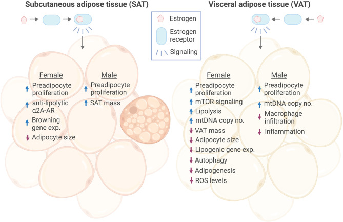
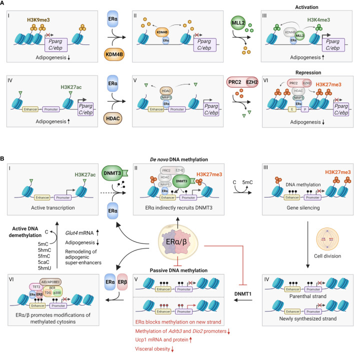
Sex hormones contribute to differences between males and females in body fat distribution and associated disease risk. Higher concentrations of estrogens are associated with a more gynoid body shape and with more fat storage on hips and thighs rather than in visceral depots. Estrogen-mediated protection against visceral adiposity is shown in post-menopausal women with lower levels of estrogens and the reduction in central body fat observed after treatment with hormone-replacement therapy. Estrogen exerts its physiological effects via the estrogen receptors (ERα, ERβ and GPR30) in target cells, including adipocytes. Studies in mice indicate that estrogen protects against adipose inflammation and fibrosis also before the onset of obesity. The mechanisms involved in estrogen-dependent body fat distribution are incompletely understood, but involve, e.g., increased mTOR signaling and suppression of autophagy and adipogenesis/lipid storage. Estrogen plays a key role in epigenetic regulation of adipogenic genes by interacting with enzymes that remodel DNA methylation and histone tail post-translational modifications. However, more studies are needed to map the differential epigenetic effects of ER in different adipocyte subtypes, including those in subcutaneous and visceral adipose tissues. We here review recent discoveries of ER-mediated transcriptional and epigenetic regulation in adipocytes, which may explain sexual dimorphisms in body fat distribution and obesity-related disease risk.

Keywords: adipocyte; epigenetic; estrogen; sexual dimorphism; steroids.


Conflict of interest statement

The authors declare that the research was conducted in the absence of any commercial or financial relationships that could be construed as a potential conflict of interest.

Figures

Figure 1
Figure 1
Effects of estrogen signaling in female and male SAT and VAT. Estrogen signaling in subcutaneous adipose tissue (SAT) and visceral adipose tissue (VAT) of both sexes has been found to promote increased proliferation of preadipocytes. Estrogen has been shown to promote anti-lipolytic effects through increasing the expression of a2A-AR in female SAT, which may, at least in part, explain the concomitant increase in SAT mass and overall anti-obesogenic effect of estrogen. In addition, Estrogen induced expression of several browning genes in female SAT. In response to estrogen, female VAT showed increased lipolysis, while lipogenic gene expression was decreased, together resulting in reduced VAT mass. On the contrary, estrogen increased male SAT volume. Adipocyte size was reduced in both female SAT and VAT by estrogen, while there were no reports of this in males. However, estrogen decreased macrophage infiltration and inflammation in male VAT. Female VAT has been shown to have reduced autophagy, adipogenesis and ROS levels in response to estrogen treatment. ER, estrogen receptor; a2A-AR, alpha2A-adrenergic receptors; exp., expression. Figure created in BioRender.com.
Figure 2
Figure 2
Epigenetic effects of ERα/β in adipocytes. (A) ERα can bind to promoter regions with repressive H3K9me3 marks (I-VI), where it recruits the histone demethylase KDM4B (also known as JMJD2B), which specifically removes these marks (II). This enables the recruitment and activity of the methyl transferase MLL2, which trimethylates lysine 4 on histone 3, forming activating H3K4me3 marks, which promotes gene expression (III). This process may occur on the promoters of Pparg and C/ebp, promoting adipogenesis. Conversely, ERα can also bind to actively transcribed genes characterized by H3K27ac marks (IV), where it binds various coregulators, including NRIP1, that enables binding of histone deacetylases (HDACs), which remove the acetyl groups on H3K27 (V). Finally, the ERα/NRIP1/HDAC complex can further bind the PRC2/EZH2 polycomb complex, which adds methyl groups to form repressive H3K27me3 marks (VI). This process can also occur on the Pparg and C/ebp promoter/enhancers, inhibiting adipogenesis. Although the repressive pathway appears most predominant, further studies should investigate whether the activating pathway indeed plays a role in certain preadipocyte/mesenchymal stem cell subpopulations. (B) ERα and ERβ affects DNA methylation through several mechanisms. ERα promotes de novo methylation and gene silencing by binding to actively transcribed regions (I), where the ERα/HDAC/PRC2/EZH2 complex first converts activating H3K27ac marks to repressive H3K27me3 marks (see Figure 2A IV-VI for details). The DNA methyl transferase DNMT3 recognizes the H3K27me3 marks, and stabilized by the ERα/HDAC/PRC2/EZH2 complex it adds a methyl group to cytosine residues on the surrounding DNA, leading to stable gene silencing (II-III). Conversely, ERα can inhibit passive DNA methylation after cell division. This occurs by transcriptional inhibition of DNMT1, which copies the DNA methylation pattern of the old DNA strand onto the newly synthesized DNA (IV-V). Red methyl groups (bottom panel V) represent hypomethylated regions in response to ERα-mediated repression of DNMT1, leading to increased beiging. ERα and/or ERβ can also promote active demethylation by recruitment of TET2, AID/APOBEC/BER complexes, which alter methylated cytosines in numerous ways that ultimately restores unmodified cytosine (VI-I). Active demethylation likely remodels adipogenic super-enhancers, and has been found to inhibit adipogenesis and increase Glut4 expression. C, Cytosine; 5mC, 5-methylcytosine; 5hmC, 5-hydroxymethylcytosine; 5fmC, 5-formylcytosine; 5caC, 5-carboxylcytosine; 5hmU, 5-hydroxymethyluracyl. Figure created in BioRender.com.

References

    1. Wells J. Sexual Dimorphism of Body Composition. Best Pract Res Clin Endocrinol Metab (2007) 21:415–30. doi: 10.1016/J.BEEM.2007.04.007 - DOI - PubMed
    1. Goossens GH, Jocken JWE, Blaak EE. Sexual Dimorphism in Cardiometabolic Health: The Role of Adipose Tissue, Muscle and Liver. Nat Rev Endocrinol (2021) 17:47–66. doi: 10.1038/S41574-020-00431-8 - DOI - PubMed
    1. Vague J. Sexual Differentiation. A Determinant Factor of the Forms of Obesity. 1947. Obes Res (1996) 4:201–3. doi: 10.1002/J.1550-8528.1996.TB00535.X - DOI - PubMed
    1. Despres JP, Nadeau A, Tremblay A, Ferland M, Moorjani S, Lupien PJ, et al. Role of Deep Abdominal Fat in the Association Between Regional Adipose Tissue Distribution and Glucose Tolerance in Obese Women. Diabetes (1989) 38:304–9. doi: 10.2337/diabetes.38.3.304 - DOI - PubMed
    1. Pouliot MC, Després JP, Nadeau A, Moorjani S, Prud’Homme D, Lupien PJ, et al. Visceral Obesity in Men: Associations With Glucose Tolerance, Plasma Insulin, and Lipoprotein Levels. Diabetes (1992) 41:826–34. doi: 10.2337/diab.41.7.826 - DOI - PubMed

Publication types