. 2022 Feb 22:10:805996.
doi: 10.3389/fbioe.2022.805996. eCollection 2022.

Efficient Transient Expression of Plasmid DNA Using Poly (2-(N, N-Dimethylamino) Ethyl Methacrylate) in Plant Cells

Affiliations

Efficient Transient Expression of Plasmid DNA Using Poly (2-(N, N-Dimethylamino) Ethyl Methacrylate) in Plant Cells

Zishuai An et al. Front Bioeng Biotechnol. .

Abstract

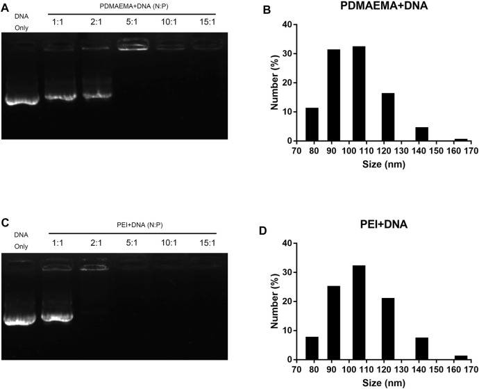
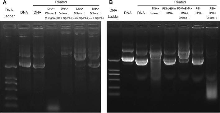
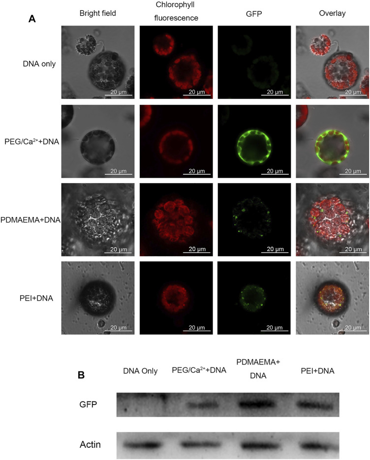
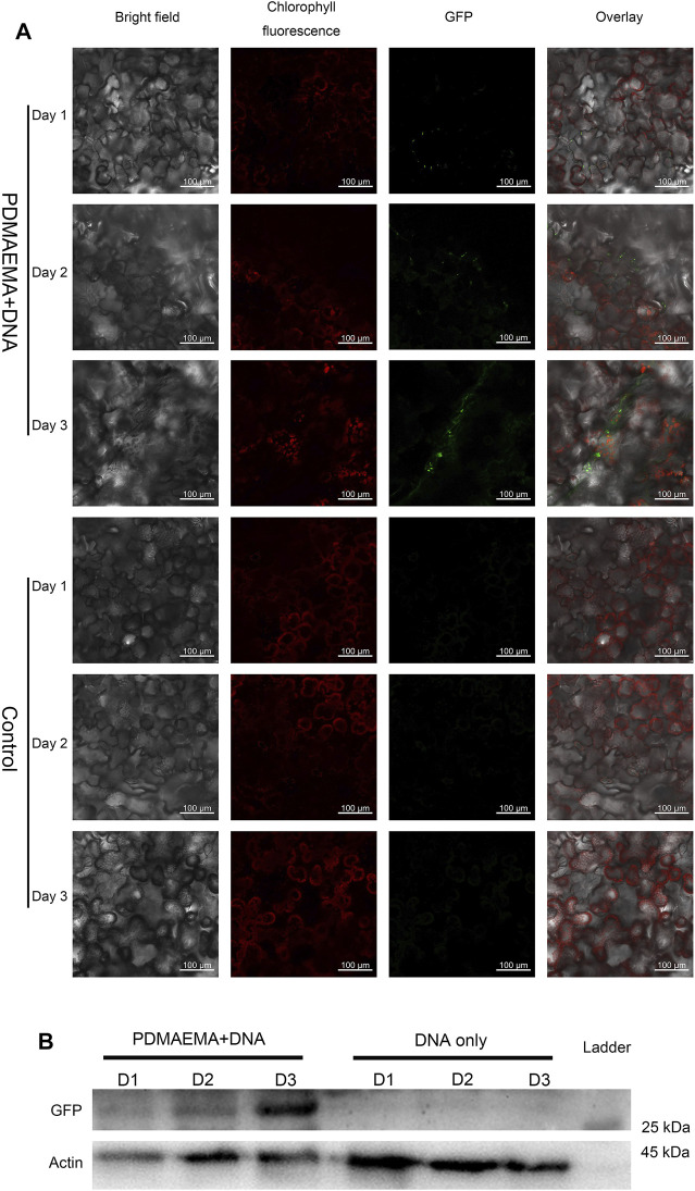
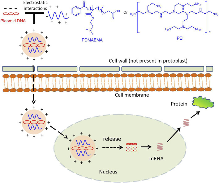
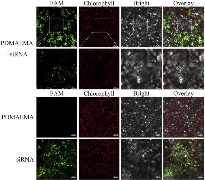
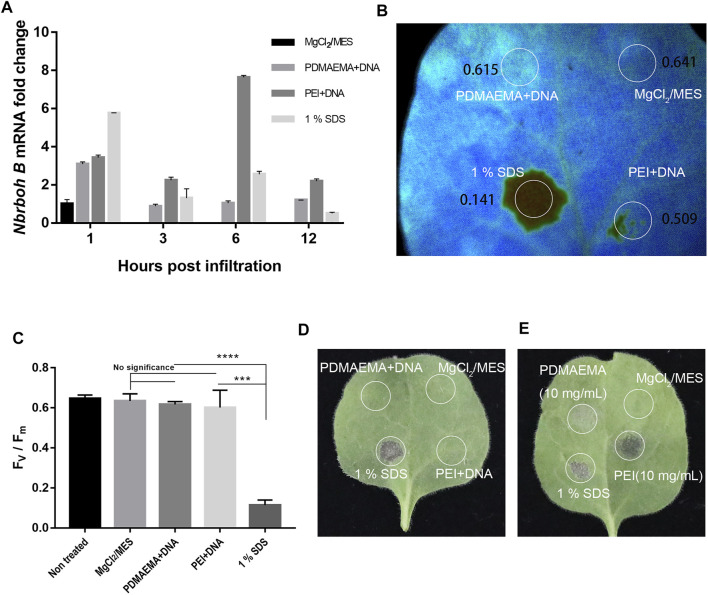
Nanomaterials have been widely studied for their potential to become the new generation of nanocarriers in gene transfection, yet it remains still difficult to apply them efficiently and succinctly to plant cells. Poly (2-(N,N-dimethylamino) ethyl methacrylate) (PDMAEMA), which possesses temperature and pH dual-sensitivity, has largely been applied in animal cells, but it is rarely involved in plant cells. As a proof of concept, PDMAEMA as a gene carrier is incubated with plasmid GFP (pGFP) to explore its transfection ability in plants, and cationic polymer polyethylenimine (PEI) is used as a control. pGFP was efficiently condensed into the nanostructure by electrostatic interactions at an N/P (amino group from cationic polymers/phosphate group from plasmid DNA (pDNA)) ratio of 15; after complexation into nanocarriers, pGFP was protected from endonuclease degradation according to the DNase I digestion assay. After incubation with protoplasts and leaves, GFP was observed with confocal microscopy in plant cells. Western blot experiments confirmed GFP expression at the protein level. Toxicity assay showed PDMAEMA had a lower toxicity than PEI. These results showed that transient expression of pGFP was readily achieved in Arabidopsis thaliana and Nicotiana benthamiana. Notably, PDMAEMA showed lower cytotoxicity than PEI upon incubation with Nicotiana benthamiana leaves. PDMAEMA exhibited great potency for DNA delivery in plant cells. This work provides us with new ideas of more concise and more effective methods for plant transformation.

Keywords: N-dimethylamino) ethyl methacrylate) (PDMAEMA); gene delivery; gene transfection; plant cells; poly (2-(N; polyethylenimine (PEI).


Conflict of interest statement

The authors declare that the research was conducted in the absence of any commercial or financial relationships that could be construed as a potential conflict of interest.

Figures

FIGURE 1
FIGURE 1
Characterization of PDMAEMA + DNA and PEI + DNA. (A) Gel retardation assay of PDMAEMA + DNA (1 μg) at N/P ratios of 1:1, 2:1, 5:1, 10:1, and 15:1. (B) Hydrodynamic diameter distributions of PDMAEMA + DNA (5 μg) (N/P ratio 15) determined by dynamic light scattering. (C) Gel retardation assay of PEI + DNA (1 μg) at N/P ratios of 1:1, 2:1, 5:1, 10:1, and 15:1. (D) Hydrodynamic diameter distributions of PEI + DNA (5 μg) (N/P ratio 15) determined by dynamic light scattering.
FIGURE 2
FIGURE 2
Plasmid DNA protection assay. (A) Same amount (1 μg) of pDNA treated by DNase I was run on a 1% agarose gel to determine the optimal DNase I concentration for DNA degradation. (B) Agarose gel electrophoresis of DNA, PDMAEMA + DNA (N/P ratio of 4.38:1), and PEI + DNA (N/P ratio of 2.6) incubated with DNase I to evaluate DNA protection against nuclease degradation.
FIGURE 3
FIGURE 3
GFP expression imaging in Arabidopsis thaliana protoplast. (A) Confocal microscope was used to image the Arabidopsis thaliana protoplast incubated with DNA only as a negative control, PEG/Ca2+ as a positive control, PDMAEMA + DNA (N/P ratio of 15:1), and PEI + DNA (N/P ratio of 15:1). GFP fluorescence images were captured with 488 nm laser excitation and 539 nm emission wavelengths. Chlorophyll autofluorescence images were captured with 633 nm laser excitation and 691 nm emission wavelengths. Scale bar, 20 μm. Protoplasts were incubated with PEG/Ca2+ for 15 min and washed with W5 solution to remove PEG. Then, protoplasts were resuspended with W5 solution in a 1.5-mL tube for another 12 h in the dark. The protoplasts were incubated with PDMAEMA + DNA or PEI + DNA directly for 12 h. (B) Western blot of the GFP expression level of Arabidopsis thaliana protoplasts incubated with DNA only, DNA-PEG/Ca2+, PDMAEMA + DNA, and PEI + DNA.
FIGURE 4
FIGURE 4
GFP expression in Nicotiana benthamiana leaves. (A) Nicotiana benthamiana leaves infiltrated with DNA only (in 10 mM MgCl2/MES as a control) and PDMAEMA + DNA (N/P ratio of 15:1) are imaged using a confocal microscope to detect GFP expression in the leaf lamina in 1 day, 2 days, and 3 days. Experiments were performed with intact leaves from healthy plants. Scale bar, 100 μm. (B) Western blot of GFP expression of Nicotiana benthamiana leaves infiltrated with DNA only (in 10 mM MgCl2/MES as control) and PDMAEMA + DNA in 1 day, 2 days, and 3 days.
FIGURE 5
FIGURE 5
Schematic illustration for the polymer-based gene delivery system in the plant cell.
SCHEME 1
SCHEME 1
PDMAEMA + FAM-siRNA NPR1 -infiltrating Nicotiana benthamiana leaves. FAM-siRNA NPR1 (20 μM) was mixed with PDMAEMA (1 mg/mL) at the fixed N/P ratio of 15 and incubated at room temperature for 30 min. The formed complexes were diluted 10 times in 10 mM MgCl2/MES (pH 5.7) before infiltration, taking only infiltrating PDMAEMA or FAM-siRNANPR1 as a control.
FIGURE 6
FIGURE 6
Toxicity assay. (A) qPCR analysis of Nbrboh B in Nicotiana benthamiana leaves with different infiltrations (MgCl2/MES (10 mM) as control, PDMAEMA + DNA (N/P ratio of 15:1), PEI + DNA (N/P ratio of 15:1), and 1% SDS at a time course. (B) Photosystem II Fv/Fm of different locations treated with PDMAEMA + DNA (N/P ratio of 15:1), PEI + DNA (N/P ratio of 15:1), 1% SDS, and MgCl2/MES (10 mM) as control on the same Nicotiana benthamiana leaf. (C) Statistical data of the (B) Fv/Fm ratio represent the variable/maximum fluorescence measurement of the photosystem II quantum efficiency. (D) Phenotype of the single Nicotiana benthamiana leaf infiltrated with MgCl2/MES (10 mM), PDMAEMA + DNA (N/P ratio of 15:1), PEI + DNA (N/P ratio of 15:1), and 1% SDS on different positions. (E) Phenotype of a single Nicotiana benthamiana leaf infiltrated with MgCl2/MES (10 mM), 1% SDS, PDMAEMA (10 mg/mL), and PEI (10 mg/mL) on different positions.

References

    1. Agarwal S., Zhang Y., Maji S., Greiner A. (2012). PDMAEMA Based Gene Delivery Materials. Mater. Today 15 (9), 388–393. 10.1016/S1369-7021(12)70165-7 - DOI
    1. Altpeter F., Springer N. M., Bartley L. E., Blechl A., Brutnell T. P., Citovsky V., et al. (2016). Advancing Crop Transformation in the Era of Genome Editing. Plant Cell 28 (7), tpc.00196.2016–1520. 10.1105/tpc.16.00196 - DOI - PMC - PubMed
    1. Baltes N. J., Gil-Humanes J., Voytas D. F. (2017). Genome Engineering and Agriculture: Opportunities and Challenges. Gene Editing in Plants 149, 1–26. 10.1016/bs.pmbts.2017.03.011 - DOI - PMC - PubMed
    1. Bijali J., Acharya K. (2020). Current Trends in Nano‐technological Interventions on Plant Growth and Development: a Review. IET nanobiotechnol. 14 (2), 113–119. 10.1049/iet-nbt.2019.0008 - DOI - PMC - PubMed
    1. Cao B., Lyu X., Wang C., Lu S., Xing D., Hu X. (2020). Rational Collaborative Ablation of Bacterial Biofilms Ignited by Physical Cavitation and Concurrent Deep Antibiotic Release. Biomaterials 262, 120341. 10.1016/j.biomaterials.2020.120341 - DOI - PubMed