. 2022 Feb 22:8:795665.
doi: 10.3389/fmed.2021.795665. eCollection 2021.

Supramaximal Horizontal Rectus Recession-Resection Surgery for Complete Unilateral Abducens Nerve Palsy

Affiliations

Supramaximal Horizontal Rectus Recession-Resection Surgery for Complete Unilateral Abducens Nerve Palsy

Zhonghao Wang et al. Front Med (Lausanne). .

Abstract

Purpose: To review the surgical procedures and outcomes of supramaximal horizontal rectus recession-resection surgery for abduction deficiency and esotropia resulting from complete unilateral abducens nerve palsy.

Methods: A total of 36 consecutive cases diagnosed as complete abducens nerve palsy, receiving supramaximal medial rectus recession (8.5 ± 1.4 mm, range: 6-10) combined with a supramaximal lateral rectus resection (11.1 ± 1.7 mm, range: 8-14) as performed over the period from 2017 to 2020, were reviewed retrospectively. All surgeries were performed by a single surgeon. Pre- and post-operative ocular motility, ocular alignment, forced duction test, binocular vision, abnormal head posture, and surgical complications were assessed.

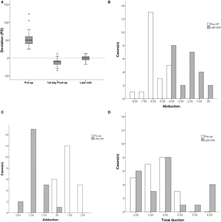
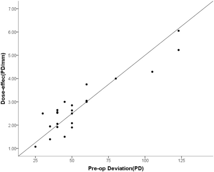
Results: Of these 36 cases, 23 (63.8%) were followed up for greater than 2 months (Mean ± SD = 8.4 ± 6.0, range: 2-24) after surgery and the collected data was presented. Mean ± SD age of these patients was 41.7 ± 14.4 (range: 12-67) years with 73.9% being female. Trauma (52.2%, 12/23) and cerebral lesions (21.7%, 5/23) were the primary etiologies for this condition. Esodeviation in primary position improved from 55.5 ± 27.2 prism diopters (PD) (range: +25 to +123) to 0.04 ± 7.3 PD (range: -18 to +12) as assessed on their last visit. Pre-operative abduction deficits of -5.6 ± 1.0 (range: -8 to -4) reduced to -2.4 ± 1.4 (range: -4 to 0) post-operatively. The mean dose-effect coefficient of 2.80 ± 1.20 PD/mm (range: 1.07-6.05) was positively correlated with pre-operative esodeviation. Rates of overcorrection and ortho were 69.6 and 26.1%, respectively, on the first day after surgery, while on their last visit the respective levels were 4.3 and 82.6%.

Conclusion: Supramaximal horizontal rectus recession-resection surgery is an effective treatment method for complete abduction deficiency. The dose-effect was positively correlated with pre-operative esodeviation. Overcorrection on the first day post-operatively is required for a long-term satisfactory surgical outcome.

Keywords: complete abducens palsy; paralytic strabismus; rectus recession-resection; surgical dosage-effect; surgical outcome.


Conflict of interest statement

The authors declare that the research was conducted in the absence of any commercial or financial relationships that could be construed as a potential conflict of interest.

Figures

Figure 1
Figure 1
Comparison of deviation (PD) and ocular motility between pre- and postoperation of patients with unilateral complete abducens nerve plasy. (A) The mean pre-operative horizontal esodeviation of 55.5 ± 27.2 PD (range: +25 to +123) significantly improved to −11.5 ± 11.0 PD (range: −35 to +12) on the first day after surgery, and further improved to 0.04 ± 7.3 PD (range: −18 to +12) as determined on their last visit. (B) The mean abduction function increased form −5.6 ± 1.0 (range: −8 to −4) preoperatively to −2.4 ± 1.4 (range: −4 to 0) on their last visit. (C) The mean adductions of the paretic eye decreased from 0.0 ± 0.0 (range: 0 to +2) pre-operatively to a mean deficiency of −1.8 ± 0.7 (range: −3 to 0) on their last visit. (D) The mean duction ranges in the paretic eye increased from 2.4 ± 1.0 pre-operatively to 3.8 ± 1.4 on their last visit. In the box plot (A), the value that is more than 1.5 times the distance from the quartile value is the outlier and is represented by “o”, while the value that is more than 2 times is the extreme value and is marked by “*”. Pre-op, pre-operatively.
Figure 2
Figure 2
The dose-effect coefficient was linearly related to the pre-operative esodeviation.
Figure 3
Figure 3
Typical case 1 showed the patient with abduction = −4 on the last visit. A 24-year-old male whose left eye deviated inward for 10 months after an injury resulting from a fall. He was diagnosed as a complete traumatic abducens nerve palsy and received a medial rectus recession of 10 mm and lateral rectus resection of 13 mm in his left eye while under general anesthesia. The FDT performed during surgery was negative. (A) The patient's 9-gaze eye position photo is shown preoperatively with left eye esotropia 50PD (LET = 50PD), abduction-6, adduction +1. (B) 1 day postoperatively with left eye exotropia 30PD (LXT = 30PD), abduction-2, adduction-4. (C) 2 months postoperatively with esotropia 3PD (ET = 3PD), abduction-4, adduction-2.
Figure 4
Figure 4
Typical case 2 showed the patient with abduction = −2 on the last visit. A 55-year-old male whose right eye deviated inward for one year after resection of a trigeminal neuroma. He was diagnosed as complete traumatic abducens nerve palsy and received a medial rectus recession of 8 mm and lateral resection of 11 mm in his right eye while under general anesthesia. The FDT performed during surgery was positive. (A) The patient's 9-gaze eye position photo is shown preoperatively with right eye esotropia 40PD (RET = 40PD), abduction-4, adduction normal. (B) 1 day postoperatively with right eye exotropia 20PD (RXT = 20 PD), abduction-1, adduction-4. (C) 2 months postoperatively with right eye exotropia 8PD (RXT = 8 PD), abduction-2, adduction-2.
Figure 5
Figure 5
Typical case 3 showed the patient with abduction = 0 on the last visit. A 47-year-old male whose right eye deviated inward for 6 years. He was diagnosed as complete traumatic abducens nerve palsy and received a medial rectus recession of 10 mm and lateral rectus resection of 12 mm in his right eye while under general anesthesia. The FDT performed during surgery was (+++). (A) The patient's 9-gaze eye position photo is shown preoperatively with right eye esotropia 60PD (RET = 60PD), abduction-5, adduction + 1.5. (B) 1 day postoperatively with right eye exotropia 20PD (RXT = 20PD), abduction 0, adduction-4. (C) 2 months post-opeatively with right eye exotropia 7PD (RXT = 7PD), abduction 0, adduction-2.

References

    1. Holmes JM, Beck RW, Kip KE, Droste PJ, Leske DA, Pediatric eye disease investigator group. predictors of non-recovery in acute traumatic sixth nerve palsy and paresis. Ophthalmology. (2001) 108:1457–60. 10.1016/S0161-6420 (01)00633-9 - DOI - PubMed
    1. Gunton KB, Medow NB, Wang FM, Nelson LB. Treatment of sixth nerve palsy. J Pediatr Ophthalmol Strabismus. (2015) 52:198–201. 10.3928/01913913-20150623-03 - DOI - PubMed
    1. Riordan-Eva P, Lee JP. Management of VIth nerve palsy–avoiding unnecessary surgery. Eye (Lond). (1992) 6:386–90. 10.1038/eye.1992.79 - DOI - PubMed
    1. Louis CV. Binocular Vision Ocular Motility. (2002), 446–8.
    1. Flanders M, Qahtani F, Gans M, Beneish R. Vertical rectus muscle transposition and botulinum toxin for complete sixth nerve palsy. Can J Ophthalmol. (2001) 36:18–25. 10.1016/S0008-4182 (01)80062-4 - DOI - PubMed

LinkOut - more resources