. 2022 Mar 2;6(1):010903.
doi: 10.1063/5.0076635. eCollection 2022 Mar.

Principles for the design of multicellular engineered living systems

Affiliations

Principles for the design of multicellular engineered living systems

Onur Aydin et al. APL Bioeng. .

Abstract

Remarkable progress in bioengineering over the past two decades has enabled the formulation of fundamental design principles for a variety of medical and non-medical applications. These advancements have laid the foundation for building multicellular engineered living systems (M-CELS) from biological parts, forming functional modules integrated into living machines. These cognizant design principles for living systems encompass novel genetic circuit manipulation, self-assembly, cell-cell/matrix communication, and artificial tissues/organs enabled through systems biology, bioinformatics, computational biology, genetic engineering, and microfluidics. Here, we introduce design principles and a blueprint for forward production of robust and standardized M-CELS, which may undergo variable reiterations through the classic design-build-test-debug cycle. This Review provides practical and theoretical frameworks to forward-design, control, and optimize novel M-CELS. Potential applications include biopharmaceuticals, bioreactor factories, biofuels, environmental bioremediation, cellular computing, biohybrid digital technology, and experimental investigations into mechanisms of multicellular organisms normally hidden inside the "black box" of living cells.


Figures

FIG. 1.
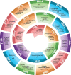
FIG. 1.
Evolutionary timeline of synthetic biology lying the foundation for M-CELS. (1962) Frog nuclei from intestinal somatic cells are re-programmed to recapitulate embryogenesis into adult frog; (1964) cracking the triplet codon genetic code; and (1970) discovery of DNA slicing/dicing with restriction endonucleases; laid foundation for recombinant DNA and genetic editing (1975) high-performance DNA sequencing; along with (1978) site-directed mutagenesis provided tools for fine dissection of gene function analysis; (1979) tissue-like architecture composed of fibroblast-synthesized collagen lattices provided framework for artificial organs; (1980) DNA cloning resulted in recombinant DNA and the birth of the biotechnology industry; (1982) discovery of the lambda phage lytic-lysogeny switch generated study of biologic circuits and systems biology; (1983) PCR amplification yielded working quantities of DNA for genomics from picograms; (1995) proteins were used as computational elements in living cells; (1995) electric circuits simulate genetic networks through hybrid modeling; (1997) artificial skin used to treat severe burn victims; (1997) Dolly, the sheep, was first mammal cloned from adult somatic cell through nuclear transfer; (1998) RNAi discovered as a tool for selective gene expression; (2000) genetic toggle switch designed in E. coli; synthetic oscillatory network designed utilizing cellular transcriptional regulators; (2001) cell–cell communication effectuates quorum sensing; (2001) Human Genome Project produces first map of the human genome; (2002) engineered gene circuits amenable to mathematical modeling; (2002) engineering and device physics of cellular logic gates; (2002) combinatorial synthesis of genetic circuits facilitates quantitative analysis of modules and systems; (2002) chemical synthesis of poliovirus cDNA de novo; (2003) design of genetic circuitry demonstrating oscillatory or toggle switch behavior; (2003) idempotent vector design for standard assembly of BioBricks; (2003) engineering the mevalonate pathway for production of terpenoids; (2004) population control circuit effected through cell–cell communication and quorum sensing; (2004) design of riboregulators facilitate posttranscriptional regulation of gene expression; (2005) genetically engineered multicellular pattern formation; (2005) design of light-sensing E. coli; (2006) induction of mouse iPSCs into specialized cells via defined factors; (2007) universal RNAi-based logic evaluator in mammalian cells; (2007) tunable mammalian genetic switch coupling repressor proteins with an RNAi target design; (2008) tunable synthetic gene oscillator; (2008) synthetic RNA devices for higher-order cellular information processing; (2009) use of multiplex genome engineering and accelerated evolution for programming cells; (2009) synthetic bacterial edge detection program; (2009) synthetic gene networks which count numerically; (2010) control of bacterial cell by genome chemically synthesized de novo; (2011) differentiation of functional human hepatocytes from iPSCs; (2011) multicellular computation via genetically encoded NOR gates; (2011) stripe patterns produced by synthetic genetic circuit; (2011) metabolic genetic switchboard designed on riboregulators; (2011) creation of sensing array from coupled biopixels; (2012) discovery of CRISPR-Cas9 as gene editing tool; (2013) generation of human cerebral organoids from iPSCs; (2013) integrating logic and memory with synthetic circuits in living cells; (2014) complete synthesis of functional eukaryotic chromosome; (2016) optogenetic control of spinal motorneuron-skeletal muscle contractility unit.
FIG. 2.
FIG. 2.
Pattern formation and self-organization in M-CELS. (a) Concentration gradients of morphogens can be generated externally or be formed in the presence of a localized source and are transported across the tissue with cell internalization acting as a sink. These morphogen gradients establish spatial patterns of cell types by regulating developmental programs. This strategy for patterning M-CELS usually results in the definition of a relatively small number of distinct regions. Inductive signals provide the patterning guide for the cells to autonomously self-organize into the relevant functional units. (b) Multiple SynNotch receptors can generate multi-layered self-organizing epithelial patterns. The epithelial layer of sender cells and a clonal population of receiver cells are co-cultivated at 1:50 ratio for 10 (start), 34 (day 1), and 58 h (day 2). Reprinted with permission from Morsut et al., Cell 164(4), 780–791 (2016). Copyright 2016 Elsevier. (c) The stochastic symmetry-breaking process occurs within the homogeneous cell mass through stochastic activation of genes leading to differentiation and cell specialization yielding cell subpopulations within the larger cell mass. (d) Specific transcription and growth factors can control lineage-specific differentiation from stem cells in M-CELS.
FIG. 3.
FIG. 3.
Development of integrated M-CELS involves interactions among multiple cellular modules. (a) Muscle-endothelium co-culture model reveals the roles of reciprocal inductive cues in angiogenesis and myogenesis. Muscle-secreted factors facilitate angiogenic sprouting, and endothelial cell-secreted factors enhance myogenic maturation. Reprinted with permission from Osaki et al., Biomaterials 156, 65–76 (2018). Copyright 2018 Elsevier. (b) Role of mechanical cues within the developmental timeline of myotendinous junctions. The formation of force-resistant attachments between myotubes and preliminary tendon structures precedes and is necessary for muscle fiber maturation. Reprinted with permission from Weitkunat et al., Current Biol. 24(7), 705–716 (2014). Copyright 2014 Elsevier. (c) Microfluidic device designs enable compartmentalization of cellular modules. In vitro neuromuscular units are developed by housing neurons and muscles in separate microfluidic channels, connected by ECM hydrogel. Reproduced with permission from Uzel et al., Sci. Adv. 2(8), e1501429 (2016Copyright 2016 Author(s), licensed under a Creative Commons Attribution 4.0 License. (d) Compartmentalized neuromuscular units are used as an in vitro model of ALS by developing co-cultures from patient-derived cells. Reproduced with permission from Osaki et al., Sci. Adv. 4(10), eaat5847 (2018). Copyright 2018 Author(s), licensed under a Creative Commons Attribution 4.0 License.
FIG. 4.
FIG. 4.
Compartmentalization and microenvironment in M-CELS' designs integrating multiple tissue constructs and organoids. (a) Microfluidic platforms can be designed for the co-culture of different cell and tissue types in controlled microenvironments with separate nutrient delivery to each cell/tissue. Reproduced with permission from Uzel et al., Sci. Adv. 2(8), e1501429 (2016). Copyright 2016 Author(s), licensed under a Creative Commons Attribution 4.0 License. (b) Microfluidics has also provided a substantial boost for a systems-level understanding of the in vitro interaction of diverse organs-on-a-chip, paving the way for the development of a synthetic organism in toto. Reprinted with permission from Wang et al., Anal. Chem. 91(13), 8667–8675 (2019). Copyright 2019 American Chemical Society. (c) Microfluidic platforms may also enable vascularization of organoids, illustrated here by the implementation of a liver organoid embedded within a vascular bed formed by HUVECs and human lung fibroblasts. The immunostaining image shows vessels formed by cells within the liver organoid (magenta alone) integrated with and supported by the vascular network formed by HUVECs (green-magenta colocalization). Unpublished work, images courtesy of Dr. Shun Zhang and Prof. Roger D. Kamm, Massachusetts Institute of Technology, Cambridge, MA. (d) Controlled spatial organization is also possible to achieve on untethered free-standing scaffolds demonstrated here by a biobot design incorporating two separate engineered muscle tissues on a 3D-printed hydrogel scaffold, producing a robotic system capable of multi-directional locomotion. Adapted with permission from Raman et al., Nat. Protoc. 12(3), 519–533 (2017). Copyright 2017 Springer Nature.
FIG. 5.
FIG. 5.
Homeostatic maintenance and engineered repair mechanisms in M-CELS. (a) The cellular microenvironment must enable M-CELS to maintain cell identity, viability, function, and the capacity to respond to various signals that may facilitate their integration into larger systems (nutrients, growth factors, cell–cell communications, and interactions). (b) Homeostatic maintenance of the microenvironment to ensure stability of the M-CELS' phenotype with closed-feedback loops and bioreactor-like control systems. (c) M-CELS must be engineered from stem cells and immune cells to enable self-repair and autonomous response to injury. Adapted with permission from Raman et al., Adv. Healthcare Mater. 6(12), 1700030 (2017). Copyright 2017 John Wiley and Sons.
FIG. 6.
FIG. 6.
M-CELS integrate and process sensory inputs to generate functional outputs. (a) M-CELS' sensing, processing, and actuation. M-CELS can sense specific extracellular and intracellular stimuli through a variety of sensors. The signals can then be processed to recognize specific levels, patterns, and combinations of stimuli, which, in turn, controls a specific set of output responses in the form of complex biological behavior that can be used for practical applications. (b) Intercellular connectivity of cardiomyocytes arranged in a serpentine pattern on a tissue engineered ray enables a global activity pattern in response to local input. Optical stimulation is applied locally at the front of the fins, and the resulting activation signal propagates via gap junctions through the body length. Republished with permission from Park et al., Science 353(6295), 158 (2016). Copyright 2016 Author(s), licensed under a Creative Commons Attribution (CC-BY-NC-ND) License. (c) Neuronal actuation and control of muscle-powered biohybrid machines: soft robotic swimmer propelled by flagella, composed of a compliant scaffold, and engineered skeletal muscle tissue innervated by motor neurons derived from optogenetic stem cells. Adapted from Aydin et al., Proc. Natl. Acad. Sci. U. S. A. 116(40), 19841–19847 (2019). Copyright 2019 Author(s), licensed under a Creative Commons Attribution (CC-BY-NC-ND) License.

References

    1. Gurdon J. B., Dev. Biol. 4, 256–273 (1962).10.1016/0012-1606(62)90043-X - DOI - PubMed
    1. Leder P. and Nirenberg M. W., Proc. Natl. Acad. Sci. U. S. A. 52(6), 1521–1529 (1964).10.1073/pnas.52.6.1521 - DOI - PMC - PubMed
    1. Smith H. O. and Welcox K. W., J. Mol. Biol. 51(2), 379–391 (1970).10.1016/0022-2836(70)90149-X - DOI - PubMed
    1. Maxam A. M. and Gilbert W., Proc. Natl. Acad. Sci. U. S. A. 74(2), 560–564 (1977).10.1073/pnas.74.2.560 - DOI - PMC - PubMed
    1. Hutchison C. A. III, Phillips S., Edgell M. H., Gillam S., Jahnke P., and Smith M., J. Biol. Chem. 253(18), 6551–6560 (1978).10.1016/S0021-9258(19)46967-6 - DOI - PubMed