Tough and Stretchy: Mechanical Properties of the Alimentary Tract in a Fish Without a Stomach
- PMID: 35274078
- PMCID: PMC8902788
- DOI: 10.1093/iob/obac003
Tough and Stretchy: Mechanical Properties of the Alimentary Tract in a Fish Without a Stomach
Abstract
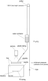
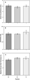
The mechanical properties of intestinal tissues determine how a thin-walled structure exerts forces on food and absorbs the force of food as it enters and travels down the gut. These properties are critically important in durophagous and stomachless fish, which must resist the potential damage to foreign bodies (e.g., shells fragments) in their diet. We test the hypothesis that the mechanical properties of the alimentary tract will differ along its length. We predict that the proximal region of the gut should be the strongest and most extensible to handle the large influx of prey often associated with stomachless fish that lack a storage depot. We developed a custom inflation technique to measure the passive mechanical properties of the whole intestine of the stomachless shiner perch, Cymatogaster aggregata. We show that mechanical properties differ significantly along the length of the alimentary tract when inflated to structural failure, with 25-46% greater maximal stress, strain, extension ratio, and toughness at the proximal (25%) position. We also find that the alimentary tissues (excluding the heavily muscular rectum) are generally highly extensible and anisotropic, and do not differ in wall circumference or thickness along the alimentary tract. These findings contribute to our knowledge of the mechanical properties of fish intestinal tissues and guide future studies of factors influencing the evolution of fish alimentary systems.
Les propriétés mécaniques des tissus intestinaux déterminent la manière dont une structure à paroi mince exerce des forces sur les aliments et absorbe leur force lorsque ceux-ci pénètrent et descendent dans l'intestin. Ces propriétés sont d'une importance capitale chez les poissons durophages et ceux sans estomac qui doivent résister aux possibles dommages que peuvent provoquer l'ingestion de corps étrangers (comme des fragments de coquilles) lorsqu'ils s'alimentent. Nous testons l'hypothèse selon laquelle les propriétés mécaniques du tube digestif diffèrent sur sa longueur. Nous prédisons que la région proximale de l'intestin devrait être la plus solide et la plus extensible pour gérer le grand afflux de proies souvent associé aux poissons sans estomac qui n'ont pas de capacité de stockage. Nous avons élaboré une technique de gonflage propre pour mesurer les propriétés mécaniques passives de l'ensemble de l'intestin de la perche méné dépourvue d'estomac, Cymatogaster aggregata. Nous montrons que les propriétés mécaniques diffèrent significativement le long du tube digestif lorsqu'il est gonflé jusqu'à une défaillance structurelle, avec une contrainte maximale, une déformation, un rapport d'extension et une résistance supérieurs de 25 à 46% à la position proximale (25%). Nous constatons également que les tissus de l'appareil digestif (à l'exclusion du rectum fortement musclé) sont généralement très extensibles et anisotropes, et ne diffèrent pas par la circonférence ou l'épaisseur de la paroi le long du tube digestif. Ces résultats contribuent à notre connaissance des propriétés mécaniques des tissus intestinaux des poissons et orientent les futures études sur les facteurs influençant l'évolution des systèmes alimentaires des poissons.
© The Author(s) 2022. Published by Oxford University Press on behalf of the Society for Integrative and Comparative Biology.
Figures


References
-
- Al-Hussaini AH. 1946. The anatomy and histology of the alimentary tract of the bottom-feeder Mulloides auriflamma (Forsk.). J Morphol 78:121–53. - PubMed
-
- Al-Hussaini AH. 1949. On the functional morphology of the alimentary tract of some fish in relation to differences in their feeding habits: anatomy and histology. Quar J of Microscopical Sci. S3:109–39. - PubMed
-
- Barrington EJW. 1957. The alimentary canal and digestion. In: The Physiology of Fish (Brown ME, ed.), p. 109–61. Academic Press, New York.
-
- Brainerd EL. 1994. Pufferfish inflation: functional morphology of postcranial structures in Diiodon holocanthus (Tetraodontiformes). J Morphol 220:243–61. - PubMed
-
- Burnstock G. 1959. The morphology of the gut of the brown trout (Salmo trutta). J of Cell Science S3-100:183–98.
LinkOut - more resources
Full Text Sources
