. 2023 Jan;41(1):142-154.
doi: 10.5534/wjmh.210177. Epub 2022 Feb 24.

Inhibition of MicroRNA-92a Improved Erectile Dysfunction in Streptozotocin-Induced Diabetic Rats via Suppressing Oxidative Stress and Endothelial Dysfunction

Affiliations

Inhibition of MicroRNA-92a Improved Erectile Dysfunction in Streptozotocin-Induced Diabetic Rats via Suppressing Oxidative Stress and Endothelial Dysfunction

Zhe Tang et al. World J Mens Health. 2023 Jan.

Abstract

Purpose: To determine whether microRNA could be a therapy target of erectile dysfunction (ED) and the underlying mechanisms.

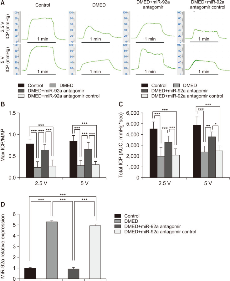
Materials and methods: Eight-week-old fasting male SD rats were intraperitoneally injected with streptozotocin to construct diabetic rat models. Diabetic ED rats were treated with miRNA-92a inhibitor. The cavernous nerves were electrically stimulated to measure the intracavernous pressure and mean arterial pressure of rats in each group. After the detection, the penile cavernous tissues are properly stored for subsequent experiments. Rat aortic endothelial cells were used in in vitro studies.

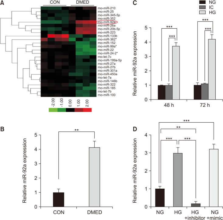
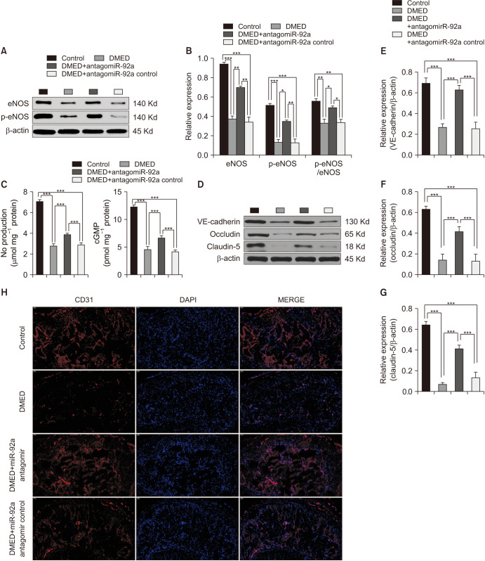
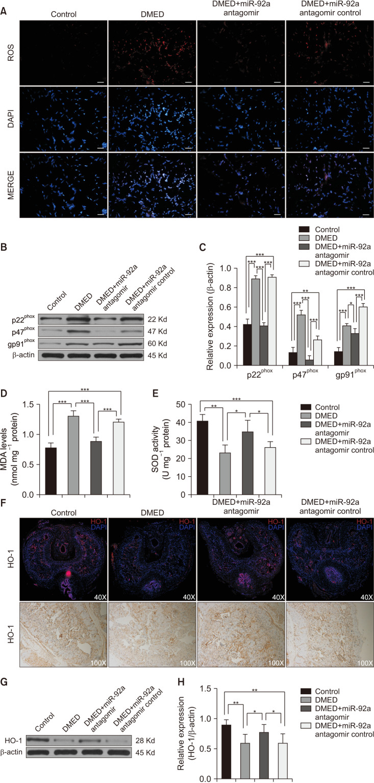
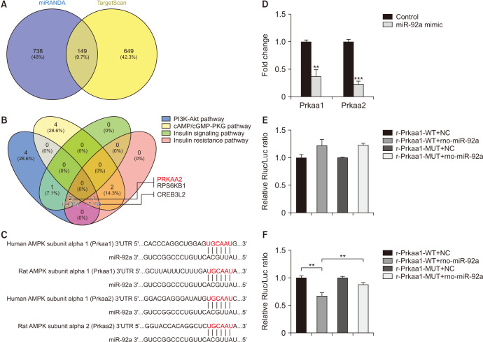
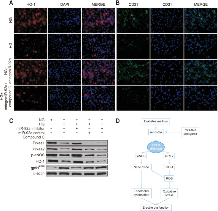
Results: The expression of miR-92a was significantly increased in the corpus cavernosum of Streptozocin (STZ)-induced diabetic rats and injection of miR-92a antagomir into the corpus cavernosum of diabetic rats significantly increased eNOS/NO/cGMP signaling pathway activities, cavernous endothelial cell proliferation, endothelial cell-cell junction protein expression and decreased the levels of oxidative stress. These changes restored erectile function in STZ-induced diabetic rats. Moreover, in vitro study demonstrated that the miR-92a expression increased significantly in endothelial cells treated with high glucose, inhibiting AMPK/eNOS and AMPK/Nrf2/HO-1 signaling pathways in rat aortic endothelial cells via targeting Prkaa2, causing endothelial dysfunction and overactive oxidative stress, miR-92a inhibitor can improve the above parameters.

Conclusions: miRNA-92a inhibitor could exert an inhibition role on oxidative stress and endothelial dysfunction to improve diabetic ED effectively.

Keywords: Endothelial cells; Erectile dysfunction; Molecular targeted therapy; Oxidative stress; microRNA.


Conflict of interest statement

The authors have nothing to disclose.

Figures

Fig. 1
Fig. 1. Elevation of miR-92a in the corpus cavernosum of DMED rats and diabetic endothelial cells. (A) Expression of miRNAs changed 3-fold above or 1/3 below in the corpus cavernosum of diabetic rats compared with CON group (n=4 per group). (B) qRT-PCR results of miR-92a expression in control group and DMED group (n=5). (C) qRT-PCR analysis showing increased expression of miR-92a in the endothelial cells treated with HG (48 h and 72 h after treatment) (n=4–5). (D) miR-92a inhibitor treatment decreased the expression of miR-92a in the endothelial cells treated with HG, while miR-92a mimic induced the expression of miR-92a in the endothelial cells treated with NG. Data are mean±SEM. **p<0.01, ***p<0.001. qRT-PCR: quantitative real-time polymerase chain reaction, CON: control, DMED: diabetic erectile dysfunction, NG: normal glucose (5 mmol/L), IC: isotonic control, HG: high glucose (30 mmol/L).
Fig. 2
Fig. 2. The miR-92a antagomir improved erectile function via decreased the expression of miR-92a in the corpus cavernosum of DMED rats. (A) Representative ICP tracing through stimulation of 2.5 volts and 5 volts for 1 minute, respective, in each group (n=10 per group). (B) The max ratio of ICP to MAP of different vols stimulation to cavernous nerve in each group (n=8–10 per group). (C) The total ICP (AUC) of different vols stimulation to cavernous nerve in each group (n=8–10 per group). (D) qRT-PCR analysis showing decreased expression of miR-92a in the corpus cavernosum of DMED rats treated with miR-92a antagomir (n=4–5 per group). Data are mean±SEM. *p<0.05, **p<0.01, ***p<0.001. DMED: diabetic erectile dysfunction, ICP: intracavernous pressure, MAP: mean arterial pressure, AUC: area under curve.
Fig. 3
Fig. 3. The miR-92a antagomir improved erectile function via amelioration of eNOS/NO/cGMP signaling pathway and endothelial cell-to-cell junctions. (A) Representative western blot results of eNOS and p-eNOS in the corpus cavernosum of all rats. (B) The expression levels of eNOS and p-eNOS, with β-actin as the loading control, and the relative ratio of p-eNOS/eNOS in the corpus cavernosum of each group are presented as bar graphs (n=6–8 per group). (C) NO and cGMP levels were determined in all four groups (n=4–5 per group). (D) Representative western blot results of VE-cadherin, occludin, and claudin-5 in the corpus cavernosum of all rats. (E-G) The expression levels of VE-cadherin, occludin, and claudin-5, with β-actin as the loading control, in the corpus cavernosum of each group are presented as bar graphs (n=6–8 per group). (H) Representative immunofluorescence result of CD31 in the corpus cavernosum of rats of all four groups. Representative immunofluorescence result of CD31 in the corpus cavernosum of rats of all four groups at ×200 magnification. Data are mean±SEM. *p<0.05, **p<0.01, ***p<0.001. DMED: diabetic erectile dysfunction, NO: nitric oxide.
Fig. 4
Fig. 4. The miR-92a antagomir improved erectile function via inhibition of oxidative stress and restoration of HO-1 expression. (A) Representative images of the ROS fluorescent probe in the corpus cavernosum of all rats at ×400 magnification. The scale bar represents 50 µm. (B, C) Representative western blot results of p22phox, p47phox, and gp91phox in the corpus cavernosum of all rats. Expressions of p22phox, p47phox, and gp91phox with β-actin as the loading control in all rats are presented as bar graphs (n=5–6 per group). (D) MDA levels determined by enzyme-linked immunosorbent assay (ELISA) in the corpus cavernosum of all rats (n=4–5 per group). (E) SOD activities determined by ELISA in the corpus cavernosum of all rats (n=4–5 per group). (F) Representative immunofluorescence and immunohistochemistry result of HO-1 in the corpus cavernosum of rats of all four groups at ×40 and ×100 magnification, respectively. (G) Representative western Blot results of HO-1 in the corpus cavernosum of all rats. (H) The expression levels of HO-1, with β-actin as the loading control, in the corpus cavernosum of each group are presented as bar graphs (n=6 per group). Data are mean±SEM. *p<0.05, **p<0.01, ***p<0.001. DMED: diabetic erectile dysfunction, ROS: reactive oxygen species, MDA: malondialdehyde, SOD: superoxide dismutase.
Fig. 5
Fig. 5. Prkaa2 was one of the genes targeted by miR-92a. (A, B) Bioinformatic analysis showing Prkaa2 was predicted as one of the genes targeted by miR-92a. (C) Predicted target region on the 3′UTR of Prkaa1 and Prkaa2 as well as the seed sequence of miR-92a by bioinformatic analysis. (D) Decreased expression of both Prkaa1 and Prkaa2 in the endothelial cells treated with miR-92a mimic. (E) Dual luciferase assay showing neither WT nor MUT Prkaa1 3′UTR activity was suppressed in the endothelial cells transfected with rno-miR-92a. (F) Dual luciferase assay showing WT but not MUT Prkaa2 3′UTR activity was suppressed in the endothelial cells transfected with rno-miR-92a. Data are mean±SEM. **p<0.01, ***p<0.001. 3′UTR: 3′ untranslated region, WT: wild type, MUT: mutant, NC: normal control.
Fig. 6
Fig. 6. HO-1 inhibition by compound C reversed the ameliorative effect of miR-92a inhibition in vitro. (A, B) Representative immunofluorescence result of HO-1 and CD31 of endothelial cells of all four groups at ×200 magnification, showing Compound C reversed the effect of miR-92a inhibitor on restoration of HO-1 and CD31 in endothelial cells. (C) Representative western blot results of Prkaa1, Prkaa2, p-eNOS, HO-1, and gp91phox of endothelial cells in each group. (D) Schematic diagram showing miR-92a-AMPK signaling axis in regulation of endothelial function and oxidative stress, and thus regulating erectile function in diabetes. NG: normal glucose, HG: high glucose, ROS: reactive oxygen species.

References

    1. Mitidieri E, Cirino G, d'Emmanuele di Villa Bianca R, Sorrentino R. Pharmacology and perspectives in erectile dysfunction in man. Pharmacol Ther. 2020;208:107493. - PubMed
    1. Ayta IA, McKinlay JB, Krane RJ. The likely worldwide increase in erectile dysfunction between 1995 and 2025 and some possible policy consequences. BJU Int. 1999;84:50–56. - PubMed
    1. McKinlay JB. The worldwide prevalence and epidemiology of erectile dysfunction. Int J Impot Res. 2000;12 Suppl 4:S6–S11. - PubMed
    1. Thorve VS, Kshirsagar AD, Vyawahare NS, Joshi VS, Ingale KG, Mohite RJ. Diabetes-induced erectile dysfunction: epidemiology, pathophysiology and management. J Diabetes Complications. 2011;25:129–136. - PubMed
    1. Ogurtsova K, da Rocha Fernandes JD, Huang Y, Linnenkamp U, Guariguata L, Cho NH, et al. IDF diabetes atlas: global estimates for the prevalence of diabetes for 2015 and 2040. Diabetes Res Clin Pract. 2017;128:40–50. - PubMed