

doi: 10.1002/cyto.a.24545.
Epub 2022 Mar 10.
OMIP 083: A 21-marker 18-color flow cytometry panel for in-depth phenotyping of human peripheral monocytes
Affiliations
- PMID: 35274803
- PMCID: PMC9310743
- DOI: 10.1002/cyto.a.24545
Item in Clipboard
OMIP 083: A 21-marker 18-color flow cytometry panel for in-depth phenotyping of human peripheral monocytes
Cytometry A.
2022 May.
No abstract available
Keywords: OMIP; PBMCs; full spectrum flow cytometry; human immunophenotyping; innate immunity; monocytes.
Conflict of interest statement
Katherine Pilkington is an employee of Cytek Biosciences, Inc., the manufacturer of the Aurora full spectrum flow cytometer used in this manuscript.
Figures
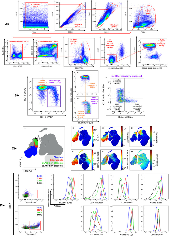
Overview of the 21‐marker, 18‐color monocyte‐centric panel on human PBMCs. PBMCs were density‐separated from Cyto‐Chex blood collection tubes (Streck, La Vista, NE), stained and acquired on a 3‐laser Cytek Aurora (Cytek biosciences, Fremont, CA). Initially, the sample is cleaned by excluding variable flow rate during acquisition (typically experienced at the start and end of sample acquisition), doublets, debris and antibody aggregates (A, i‐vi). Following cleaning, CD45+ (A, vii) and CD66b− (A, viii) leukocytes are sequentially gated. Several lineage markers (CD3, CD19, CD56, CD123) are conjugated to the same fluorochrome to facilitate efficient exclusion of all other nonmonocyte cells (A, ix). Within the same plot that Lin+ leukocytes are excluded, the monocyte population is defined as TLR2+ (A, ix). As such, the monocyte population is defined as: CD45+/CD66b−/Lin−/TLR2+. Monocytes display their characteristic expression of CD14 and CD16 (B, i). Separating classical (CD14++/CD16−) monocytes from all other monocyte subsets (CD14var/CD16var) can be achieved in this plot. From here, classical monocytes are confirmed to display their expected pattern of CCR2 and CX3CR1 (B, ii). Residual classical monocytes are defined in B, iii as those cells which were originally gated as ‘other monocyte subsets’ from the parent population but which subsequently show CCR2 and CX3CR1 expression patterns identical to classical monocytes. This population is removed from downstream analysis. From here, a true population of ‘other monocyte subsets’ is defined (B, iii) and subsequently divided into intermediate (CCR2+/SLAN−) monocytes and two subsets of nonclassical monocytes based on expression of SLAN: SLAN+ (CCR2−/SLAN+) nonclassical and SLAN− (CCR2−/SLAN−) monocytes (B, iv). High‐dimensional analysis with the UMAP algorithm was conducted on PBMCs from two individuals across three timepoints (for a total of six samples) (C). Data were cleaned (as demonstrated in A, i‐vi) and CD45+ events from these six samples were combined and used for this analysis. Manual gating of the four monocyte populations was overlaid on this two‐dimensional rendering (C, i). UMAP analysis demonstrates the ease of discriminating the monocyte population from all other leukocytes using TLR2 (C, ii). Heatmap overlays also demonstrate the distinct expression levels of CD14 (C, iii), CD16 (C, iv), CCR2 (C, v), CX3CR1 (C, vi) and SLAN (C, vii) within these monocyte populations. Once identified, these four monocyte subsets were interrogated for their expression of a further nine markers (D); each monocyte subset shows distinctive expression profiles
Similar articles
-
OMIP-069: Forty-Color Full Spectrum Flow Cytometry Panel for Deep Immunophenotyping of Major Cell Subsets in Human Peripheral Blood.Cytometry A. 2020 Oct;97(10):1044-1051. doi: 10.1002/cyto.a.24213. Epub 2020 Aug 31. Cytometry A. 2020. PMID: 32830910 Free PMC article.
-
OMIP-109: 45-color full spectrum flow cytometry panel for deep immunophenotyping of the major lineages present in human peripheral blood mononuclear cells with emphasis on the T cell memory compartment.Cytometry A. 2024 Nov;105(11):807-815. doi: 10.1002/cyto.a.24900. Epub 2024 Oct 28. Cytometry A. 2024. PMID: 39466962
-
OMIP-94: Twenty-four-color (thirty-marker) panel for deep immunophenotyping of immune cells in human peripheral blood.Cytometry A. 2023 Sep;103(9):695-702. doi: 10.1002/cyto.a.24766. Epub 2023 May 30. Cytometry A. 2023. PMID: 37254600
-
Comprehensive phenotyping of peripheral blood monocytes in healthy bovine.Cytometry A. 2022 Feb;101(2):122-130. doi: 10.1002/cyto.a.24492. Epub 2021 Aug 12. Cytometry A. 2022. PMID: 34382742 Review.
-
Flow cytometry immunophenotyping in integrated diagnostics of patients with newly diagnosed cytopenia: one tube 10-color 14-antibody screening panel and 3-tube extensive panel for detection of MDS-related features.Int J Lab Hematol. 2015 May;37 Suppl 1:133-43. doi: 10.1111/ijlh.12368. Int J Lab Hematol. 2015. PMID: 25976971 Review.
Cited by
-
Causal role of immune cells in uveitis: Mendelian randomization study.Front Immunol. 2024 Jul 9;15:1402074. doi: 10.3389/fimmu.2024.1402074. eCollection 2024. Front Immunol. 2024. PMID: 39044820 Free PMC article.
-
Single-Cell Atlas of Atherosclerosis Patients by Cytof: Circulatory and Local Immune Disorders.Aging Dis. 2024 Feb 1;15(1):245-258. doi: 10.14336/AD.2023.0426-1. Aging Dis. 2024. PMID: 37307820 Free PMC article.
-
Integrating transcriptomics, eQTL, and Mendelian randomization to dissect monocyte roles in severe COVID-19 and gout flare.Front Genet. 2024 Sep 25;15:1385316. doi: 10.3389/fgene.2024.1385316. eCollection 2024. Front Genet. 2024. PMID: 39385934 Free PMC article.
-
Peripheral monocyte subsets are altered during gestation in oocyte donation pregnancy complicated with pre-eclampsia.Scand J Immunol. 2025 Feb;101(2):e13432. doi: 10.1111/sji.13432. Scand J Immunol. 2025. PMID: 39920890 Free PMC article.
-
Design of a Robust Flow Cytometric Approach for Phenotypical and Functional Analysis of Human Monocyte Subsets in Health and Disease.Biomolecules. 2024 Oct 3;14(10):1251. doi: 10.3390/biom14101251. Biomolecules. 2024. PMID: 39456184 Free PMC article.
References
-
- Niewold P, Ashhurst TM, Smith AL, King NJC. Evaluating spectral cytometry for immune profiling in viral disease. Cytometry A. 2020;97(11):1165–79. - PubMed
-
- Bocsi J, Melzer S, Dähnert I, Tárnok A. OMIP‐023: 10‐color, 13 antibody panel for in‐depth phenotyping of human peripheral blood leukocytes. Cytometry A. 2014;85(9):781–4. - PubMed
-
- Baumgart S, Peddinghaus A, Schulte‐Wrede U, Mei HE, Grätzkau A. OMIP‐034: comprehensive immune phenotyping of human peripheral leukocytes by mass cytometry for monitoring immunomodulatory therapies. Cytometry A. 2017;91(1):34–8. - PubMed
Publication types
MeSH terms
LinkOut - more resources
Full Text Sources
