

Cortical Features in Child and Adolescent Carriers of Mutant Huntingtin (mHTT)
- PMID: 35275555
- PMCID: PMC9177765
- DOI: 10.3233/JHD-210512
Cortical Features in Child and Adolescent Carriers of Mutant Huntingtin (mHTT)
Abstract
Background: Molecular studies provide evidence that mutant huntingtin (mHTT) affects early cortical development; however, cortical development has not been evaluated in child and adolescent carriers of mHTT.
Objective: To evaluate the impact of mHTT on the developmental trajectories of cortical thickness and surface area.
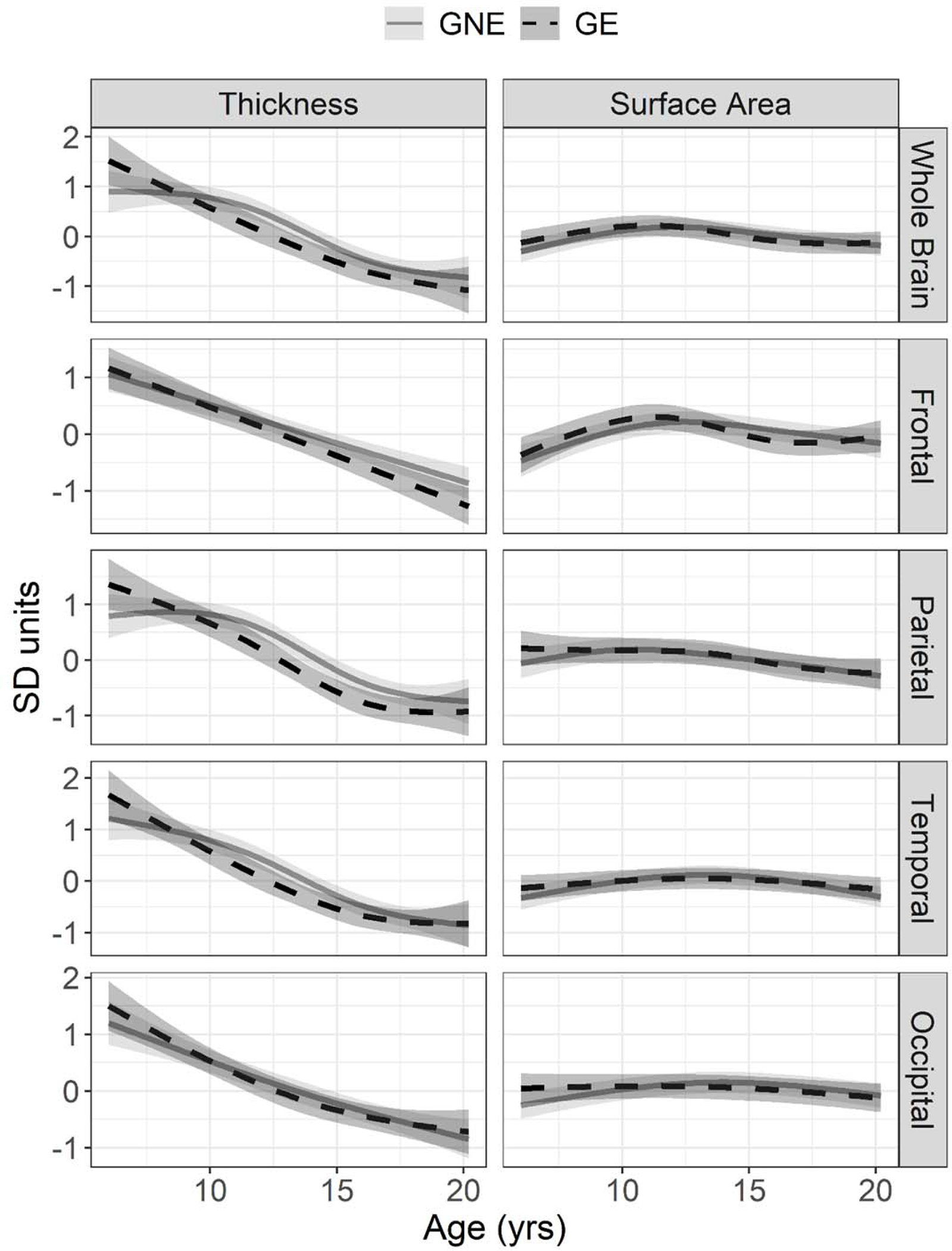
Methods: Children and adolescents (6-18 years) participated in the KidsHD study. mHTT carrier status was determined for research purposes only to classify participants as gene expanded (GE) and gene non-expanded (GNE). Cortical features were extracted from 3T neuroimaging using FreeSurfer. Nonlinear mixed effects models were conducted to determine if age, group, and CAG repeat were associated with cortical morphometry.
Results: Age-related changes in cortical morphometry were similar across groups. Expanded CAG repeat was not significantly associated with cortical features.
Conclusion: While striatal development is markedly different in GE and GNE, developmental change of the cortex appears grossly normal among child and adolescent carrier of mHTT.
Keywords: Huntington’s disease; children at risk for HD; cortical development; magnetic resonance imaging; trinucleotide repeat disorder.
Conflict of interest statement
Conflict of Interest
The authors report no other conflicts of interest relevant to this work.
Figures
References
Publication types
MeSH terms
Substances
Grants and funding
LinkOut - more resources
Full Text Sources
Medical
