. 2022 Mar 11;8(10):eabj8331.
doi: 10.1126/sciadv.abj8331. Epub 2022 Mar 11.

Platelets drive fibronectin fibrillogenesis using integrin αIIbβ3

Affiliations

Platelets drive fibronectin fibrillogenesis using integrin αIIbβ3

Sebastian Lickert et al. Sci Adv. .

Abstract

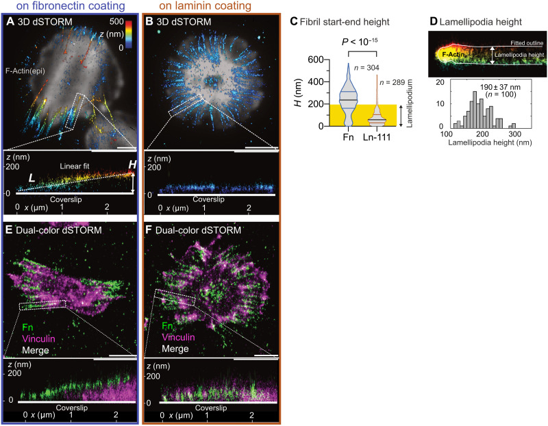
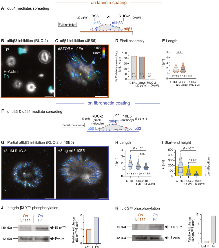
Platelets interact with multiple adhesion proteins during thrombogenesis, yet little is known about their ability to assemble fibronectin matrix. In vitro three-dimensional superresolution microscopy complemented by biophysical and biochemical methods revealed fundamental insights into how platelet contractility drives fibronectin fibrillogenesis. Platelets adhering to thrombus proteins (fibronectin and fibrin) versus basement membrane components (laminin and collagen IV) pull fibronectin fibrils along their apical membrane versus underneath their basal membrane, respectively. In contrast to other cell types, platelets assemble fibronectin nanofibrils using αIIbβ3 rather than α5β1 integrins. Apical fibrillogenesis correlated with a stronger activation of integrin-linked kinase, higher platelet traction forces, and a larger tension in fibrillar-like adhesions compared to basal fibrillogenesis. Our findings have potential implications for how mechanical thrombus integrity might be maintained during remodeling and vascular repair.


Figures

Fig. 1.
Fig. 1.. Fn extracellular matrix assembled by human platelets on Fn-coated and Ln-111–coated coverslips.
(A) Sketch of the experimental procedure. Platelets are seeded on Fn-coated or Ln-coated glass coverslips for 2 hours at 37°C in a medium supplemented with pFn (90 μg ml−1) and pFn647 (10 μg ml−1). During this time, platelets incorporate labeled pFn into newly assembled Fn fibers. Samples are then fixed and imaged by superresolution microscopy. (B) 3D STORM of pFn647 assembled by platelets on Fn-coated glass coverslips. The z position of pFn647 is color-coded from blue (basal) to red (apical). All STORM images are overlaid onto an epifluorescence (epi) image of F-actin (gray). (C) Magnified view of pFn fibrils at the cell periphery showing submicrometer-long Fn deposits. (D) Magnified view of pFn fibrils at the cell end showing micrometer-long Fn fibrils. (E) 3D STORM of Fn647 assembled by platelets on Ln-111–coated glass coverslips. (F) Magnified view of radially oriented micrometer-long Fn fibrils. (G) Length L and (H) thickness D of pFn fibrils extracted from 3D STORM data of fibrils formed on Fn versus Ln-111 coatings. Data distributions are depicted as violin plots showing the median (thick black line) and interquartile ranges (thin colored lines). Data were compared with an unpaired two-tailed Mann-Whitney test. Adjusted P < 0.001 was accepted as highly significant. n.s., not significant. Data were pooled from nine donors (24 to 35 years). Only fibrils longer than 1 μm were included in the analysis. Scale bars, 5 μm (B and E) and 1 μm (C, D, and F).
Fig. 2.
Fig. 2.. Temporal sequence of Fn fibril assembly by platelets and dependence on lamellipodia dynamics.
(A to D) 3D STORM of pFn647 assembled by platelets on Fn-coated glass coverslips after 15 (A), 30 (B), 45 (C), and 120 min (D). Shown are representative platelets for each time point. The z position of pFn647 is color-coded from blue (basal) to red (apical). All STORM images are overlaid onto an epifluorescence (epi) image of F-actin (gray). Bottom row: Magnified views of the boxed regions. (E to H) pFn647 assembled by platelets on Ln-coated glass coverslips after 15 (E), 30 (F), 45 (G), and 120 min (H). Bottom row: Magnified views of the boxed regions. (I) pFn647 assembled on Fn-coatings by platelets treated with 100 μM change to Rac-1 selective inhibitor NSC23766. Right: Magnified view. (J) Representative dual-color STORM image of pFn680 (cyan) and phalloidin-647 (gray) in a single platelet seeded for 120 min on Fn-coated glass. Platelets were obtained from three donors (24 to 34 years). Scale bars, 5 μm (I), 2 μm (A to H, top rows, and J), and 1 μm (A to I, magnified views).
Fig. 3.
Fig. 3.. Analysis of the dimensionality of Fn fibrils assembled by human platelets seeded on Fn and Ln coatings.
(A) 3D STORM of Fn fibrils assembled by a representative platelet spread on Fn-coated glass. The z position of pFn647 is color-coded from blue (basal) to red (apical). All STORM images are overlaid onto an epifluorescence (epi) image of F-actin (gray). Bottom: Side view of a single Fn fibril [boxed region in (A)] and linear fit (dashed line) yielding the length L and the height H of the fibril. (B) Fn fibril assembly by a representative platelet spread on a Ln-111–coated coverslip. Note the nonstraight appearance of Fn fibrils on Ln-111. Bottom: Side view of a single Fn fibril [boxed region in (B)]. (C) Comparison of Fn fibril height on Fn coatings (blue) and Ln-111 coatings (brown). The yellow background denotes the lamellipodia height. Data were pooled from nine donors (24 to 35 years). Only fibrils longer than 1 μm were included in the analysis; for donor-to-donor variation on Fn and Ln-111 coatings, see fig. S2. Data were compared using an unpaired two-tailed Mann-Whitney test. (D) Analysis of lamellipodia height of spread platelets on Fn (cf. fig. S8). One hundred regions in 13 spread platelets were analyzed. (E) Dual-color 3D STORM of pFn647 (green) and vinculin (magenta) in a representative platelet spread on Fn. Bottom: Side view of a single Fn fibril [boxed region in (E)]. (F) Dual-color 3D STORM of pFn647 (green) and vinculin (magenta) in a representative platelet spread on Ln. Bottom: Side view of a single Fn fibril [boxed region in (F)]. Scale bars, 2 μm (A, B, E, and F).
Fig. 4.
Fig. 4.. Contributions of α5β1 and αIIbβ3 integrins to Fn fibrillogenesis by platelets.
(A) Saturating concentrations of the blocking antibody JBS5 (20 μg ml−1) against α5β1 or of the nonpriming inhibitor RUC-2 (100 μM) against αIIbβ3 were supplemented during platelet seeding on Ln-111–coated glass coverslips. Platelets spread on Ln using α6β1 integrins independent of the blocked integrins. (B) Epifluorescence image of F-actin (gray) and pFn647 (cyan) of platelets inhibited with RUC-2 (100 μM). (C) 3D STORM of Fn fibrils (z color code) overlaid onto an epifluorescence F-actin image (gray) of a platelet inhibited with JBS5 (20 μg ml−1). (D) Fraction of platelets that produced micrometer-long fibrils in the absence or presence of JBS5 and RUC-2, respectively. (E) Fn fibril length for platelets treated with JBS5 or RUC-2. Data were obtained from three donors (28 and 34 years). No fibrils were detected for RUC-2 (n.d., not detectable). (F) Subsaturating concentrations of the nonpriming αIIbβ3 inhibitor RUC-2 (3 μM) or of the blocking antibody 10E5 (3 μg ml−1) against αIIbβ3 were supplemented during platelet seeding on Fn-coated glass coverslips. (G) 3D STORM of Fn fibrils assembled in the presence of 3 μM RUC-2 (left) or 10E5 (3 μg ml−1) (right). (H and I) Fibril dimensions for partial αIIbβ3 inhibition. Data were pooled from two donors (28 to 35 years) and compared with an unpaired two-tailed Mann-Whitney test. (J and K) Cell lysates of platelets seeded on Ln-111– or Fn-coated coverslips were separated by SDS–polyacrylamide gel electrophoresis and stained for (J) integrin β3 pY773 or (K) integrin-linked kinase (ILK) pS246. The level of protein phosphorylation was quantified and normalized by β-actin. Scale bars, 5 μm (B) and 2 μm (C and G).
Fig. 5.
Fig. 5.. Nanoscopic analysis of fibrillar-like adhesion molecular morphology.
(A) Dual-color STORM image of pFn647 (green) and vinculin (magenta) in a representative platelet spread on Fn-coated glass. Bottom: Magnified view of the boxed region in (A) along a Fn fibril. Red arrow (Δx) denotes the spatial offset of the vinculin stain with respect to the Fn fibril. (B) Representative platelet seeded in the presence of subsaturating concentrations of the αIIbβ3 blocking antibody 10E5 (3 μg ml−1) on Fn-coated glass. Same representation as in (A). (C) pFn647 (green) and vinculin (magenta) in a representative platelet spread on Ln-111–coated glass. Same representation as in (A). (D) Exemplary line profiles (dashed) of the density of fluorophore localizations along the line in (A) for the vinculin stain (magenta) and the pFn stain (green). Each profile was approximated by a step function to obtain the spatial offset Δx between stains. (E) Spatial offset between Fn fibrils and vinculin for platelets on Fn, on Ln-111, or on Fn with partial inhibition of αIIbβ3. Platelets were obtained from three donors (28 to 34 years). Data were compared using Kruskall-Wallis rank test with post hoc Dunn’s test. Scale bars, 2 μm (A to C) and 1 μm (A, magnified view).
Fig. 6.
Fig. 6.. Cell-substrate tractions of platelets on Fn and Ln.
(A) Traction forces of single platelets are measured with an optimized micropost array. The tops of the posts are stamped with the respective proteins (blue) and the side walls are passivated (green). Post deflections are determined from the centroids of posts in confocal slices using fluorescently labeled bovine serum albumin (BSA) (green). (B) Platelets are seeded for 1 hour on microposts arrays coated with Fn (left) or Ln-111 (right) and then fixed and stained for F-actin (red hot color map). Insets: Force distribution (red arrows) and measured values of a representative platelet (white boxed region). Force scale bars, 5 nN. (C to E) Comparisons between Fn and Ln-111 in terms of (C) spreading area on posts, (D) the mean force per post, and (E) total force per platelet. The total force per cell is the sum of the magnitude of forces acting on individual posts beneath a single cell, not their vectorial sum. Data were pooled from four donors (33 to 43 years). Data were compared using an unpaired two-tailed Mann-Whitney test. Scale bars, 10 μm (B) and 2 μm (A).
Fig. 7.
Fig. 7.. Fn fibrillogenesis by platelets in the context of basement membrane proteins or clot proteins.
(A) 3D STORM image of pFn647 of a single platelet seeded on Fb (binds αIIbβ3) overlaid onto an epifluorescence (epi) image of F-actin (gray). (B) 3D STORM image of pFn647 of a single platelet seeded on Col4 (binds α2β1) overlaid onto an epifluorescence (epi) image of F-actin (gray). (C) Fibril start-to-end height between Fn, Fb, Col4, and Ln-111. Data were pooled from four donors (33 to 43 years) and were compared with a nonparametric Kruskall-Wallis rank test with post hoc Dunn’s test to make multiple comparisons. (D) Platelet traction forces on Fn, Fg, Col4, and Ln-111–coated micropost arrays. (E) Epifluorescence image of F-actin (left) and 3D STORM image of pFn647 (middle) of platelet aggregates formed on Ln. Note that the color code corresponds to an extended z-range of 1.2 μm. Right: z slices showing the upper 0.8 μm (top) or lower 0.4 μm (bottom) of the STORM data (same color code). (F) Lattice light sheet micrograph of Fn (green) and Fb (magenta) in a clot formed from whole blood on a Fn-coated surface. Left: Average intensity projection of the stack. Right: Selected slices of the boxed regions in (E). Scale bars, 50 μm (F), 10 μm (E and F, insets), and 2 μm (A and B).
Fig. 8.
Fig. 8.. Integrin mechanosensing of ECM proteins triggers dimensionality of the first deposited ECM network.
Schematic summary of results. Platelets spread on Fn and Fb (right) generate high traction forces (red arrows and dark red-yellow) and Fn fibrils are aligned to polarized actin bundles (top view). Mechanosignaling (black arrow) through αIIbβ3 (blue) instructs Fn fibril anchorage along the apical membrane of platelets and the mechanomolecular strain induces a spatial offset between vinculin (green) and Fn (see inset). On Ln or Col4 (middle), platelets contract less strong (yellow) and form radial oriented Fn fibrils (top view) pulled beneath their basal side with a small spatial offset between vinculin and Fn (side view, inset). Dose-dependent inhibition of myosin IIa and the αIIbβ3 (left) further reduces platelet contractility (green) and prevents Fn fibril formation.

Similar articles

Cited by

References

    1. Rodrigues M., Kosaric N., Bonham C. A., Gurtner G. C., Wound healing: A cellular perspective. Physiol. Rev. 99, 665–706 (2019). - PMC - PubMed
    1. Ting L. H., Feghhi S., Taparia N., Smith A. O., Karchin A., Lim E., John A. S., Wang X., Rue T., White N. J., Sniadecki N. J., Contractile forces in platelet aggregates under microfluidic shear gradients reflect platelet inhibition and bleeding risk. Nat. Commun. 10, 1204 (2019). - PMC - PubMed
    1. Myers D. R., Qiu Y., Fay M. E., Tennenbaum M., Chester D., Cuadrado J., Sakurai Y., Baek J., Tran R., Ciciliano J. C., Ahn B., Mannino R. G., Bunting S. T., Bennett C., Briones M., Fernandez-Nieves A., Smith M. L., Brown A. C., Sulchek T., Lam W. A., Single-platelet nanomechanics measured by high-throughput cytometry. Nat. Mater. 16, 230–235 (2017). - PMC - PubMed
    1. Nurden P., Nurden A. T., Congenital disorders associated with platelet dysfunctions. Thromb. Haemost. 99, 253–263 (2008). - PubMed
    1. Nicolai L., Schiefelbein K., Lipsky S., Leunig A., Hoffknecht M., Pekayvaz K., Raude B., Marx C., Ehrlich A., Pircher J., Zhang Z., Saleh I., Marel A. K., Löf A., Petzold T., Lorenz M., Stark K., Pick R., Rosenberger G., Weckbach L., Uhl B., Xia S., Reichel C. A., Walzog B., Schulz C., Zheden V., Bender M., Li R., Massberg S., Gaertner F., Vascular surveillance by haptotactic blood platelets in inflammation and infection. Nat. Commun. 11, 5778 (2020). - PMC - PubMed