

Structural basis for HCMV Pentamer recognition by neuropilin 2 and neutralizing antibodies
- PMID: 35275718
- PMCID: PMC8916728
- DOI: 10.1126/sciadv.abm2546
Structural basis for HCMV Pentamer recognition by neuropilin 2 and neutralizing antibodies
Abstract
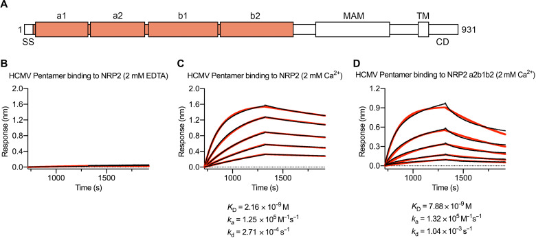
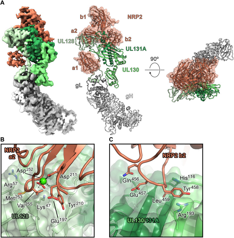
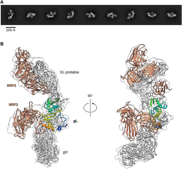
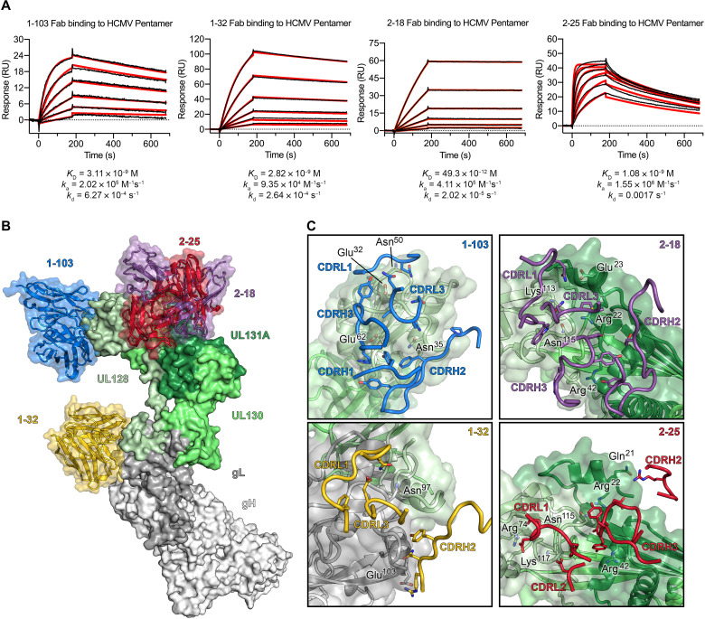
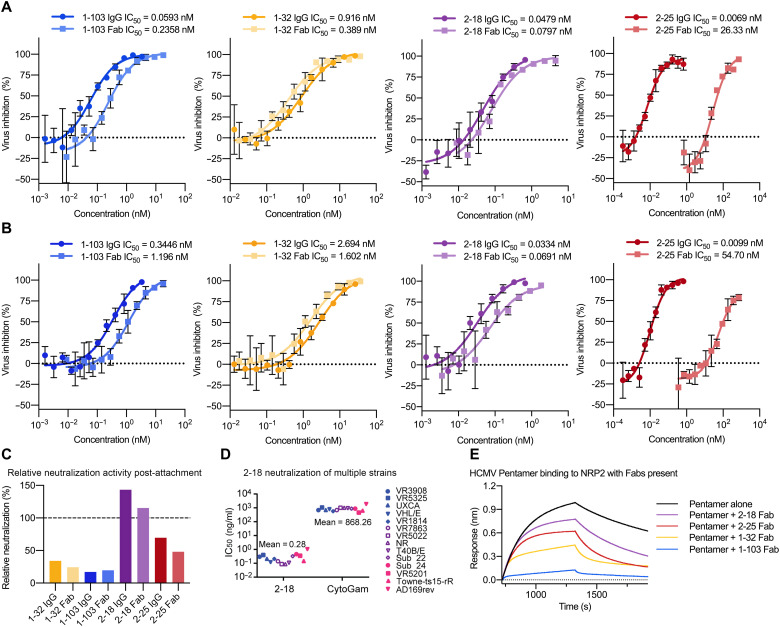
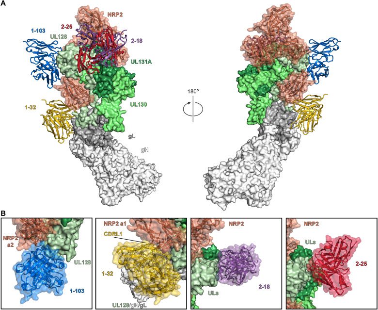
Human cytomegalovirus (HCMV) encodes multiple surface glycoprotein complexes to infect a variety of cell types. The HCMV Pentamer, composed of gH, gL, UL128, UL130, and UL131A, enhances entry into epithelial, endothelial, and myeloid cells by interacting with the cell surface receptor neuropilin 2 (NRP2). Despite the critical nature of this interaction, the molecular determinants that govern NRP2 recognition remain unclear. Here, we describe the cryo-EM structure of NRP2 bound to Pentamer. The high-affinity interaction between these proteins is calcium dependent and differs from the canonical carboxyl-terminal arginine (CendR) binding that NRP2 typically uses. We also determine the structures of four neutralizing human antibodies bound to the HCMV Pentamer to define susceptible epitopes. Two of these antibodies compete with NRP2 binding, but the two most potent antibodies recognize a previously unidentified epitope that does not overlap the NRP2-binding site. Collectively, these findings provide a structural basis for HCMV tropism and antibody-mediated neutralization.
Figures
Similar articles
-
Structural basis for HCMV Pentamer receptor recognition and antibody neutralization.Sci Adv. 2022 Mar 11;8(10):eabm2536. doi: 10.1126/sciadv.abm2536. Epub 2022 Mar 11. Sci Adv. 2022. PMID: 35275719 Free PMC article.
-
Neutralization Epitopes in Trimer and Pentamer Complexes Recognized by Potent Cytomegalovirus-Neutralizing Human Monoclonal Antibodies.Microbiol Spectr. 2022 Dec 21;10(6):e0139322. doi: 10.1128/spectrum.01393-22. Epub 2022 Nov 7. Microbiol Spectr. 2022. PMID: 36342276 Free PMC article.
-
Structural and biochemical studies of HCMV gH/gL/gO and Pentamer reveal mutually exclusive cell entry complexes.Proc Natl Acad Sci U S A. 2015 Feb 10;112(6):1767-72. doi: 10.1073/pnas.1424818112. Epub 2015 Jan 26. Proc Natl Acad Sci U S A. 2015. PMID: 25624487 Free PMC article.
-
Human cytomegalovirus tropism for endothelial/epithelial cells: scientific background and clinical implications.Rev Med Virol. 2010 May;20(3):136-55. doi: 10.1002/rmv.645. Rev Med Virol. 2010. PMID: 20084641 Review.
-
Neutralization of Human Cytomegalovirus Entry into Fibroblasts and Epithelial Cells.Vaccines (Basel). 2017 Oct 31;5(4):39. doi: 10.3390/vaccines5040039. Vaccines (Basel). 2017. PMID: 29088098 Free PMC article. Review.
Cited by
-
The human cytomegalovirus decathlon: Ten critical replication events provide opportunities for restriction.Front Cell Dev Biol. 2022 Nov 25;10:1053139. doi: 10.3389/fcell.2022.1053139. eCollection 2022. Front Cell Dev Biol. 2022. PMID: 36506089 Free PMC article. Review.
-
NRP1 is a receptor for mammalian orthoreovirus engaged by distinct capsid subunits.Cell Host Microbe. 2024 Jun 12;32(6):980-995.e9. doi: 10.1016/j.chom.2024.04.014. Epub 2024 May 9. Cell Host Microbe. 2024. PMID: 38729153 Free PMC article.
-
Discrimination of primary and chronic cytomegalovirus infection based on humoral immune profiles in pregnancy.J Clin Invest. 2024 Oct 15;134(20):e180560. doi: 10.1172/JCI180560. J Clin Invest. 2024. PMID: 39207860 Free PMC article.
-
miR-24-3p Is Antiviral Against SARS-CoV-2 by Downregulating Critical Host Entry Factors.Viruses. 2024 Nov 28;16(12):1844. doi: 10.3390/v16121844. Viruses. 2024. PMID: 39772154 Free PMC article.
-
A broadly neutralizing human monoclonal antibody generated from transgenic mice immunized with HCMV particles limits virus infection and proliferation.J Virol. 2024 Jul 23;98(7):e0021324. doi: 10.1128/jvi.00213-24. Epub 2024 Jun 4. J Virol. 2024. PMID: 38832789 Free PMC article.
References
-
- Cannon M. J., Schmid D. S., Hyde T. B., Review of cytomegalovirus seroprevalence and demographic characteristics associated with infection. Rev. Med. Virol. 20, 202–213 (2010). - PubMed
-
- Ahlfors K., Forsgren M., Ivarsson S. A., Harris S., Svanberg L., Congenital cytomegalovirus infection: On the relation between type and time of maternal infection and infant’s symptoms. Scand. J. Infect. Dis. 15, 129–138 (1983). - PubMed
-
- Pass R. F., Fowler K. B., Boppana S. B., Britt W. J., Stagno S., Congenital cytomegalovirus infection following first trimester maternal infection: Symptoms at birth and outcome. J. Clin. Virol. 35, 216–220 (2006). - PubMed
Grants and funding
LinkOut - more resources
Full Text Sources
Miscellaneous
