. 2022 Mar 11;8(10):eabn3264.
doi: 10.1126/sciadv.abn3264. Epub 2022 Mar 11.

d-Serine controls epidermal vesicle release via NMDA receptor, allowing tissue migration during the metamorphosis of the chordate Ciona

Affiliations

d-Serine controls epidermal vesicle release via NMDA receptor, allowing tissue migration during the metamorphosis of the chordate Ciona

Gabriel Krasovec et al. Sci Adv. .

Abstract

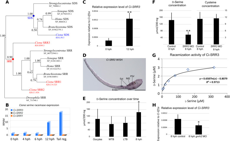
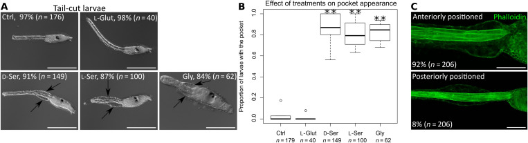
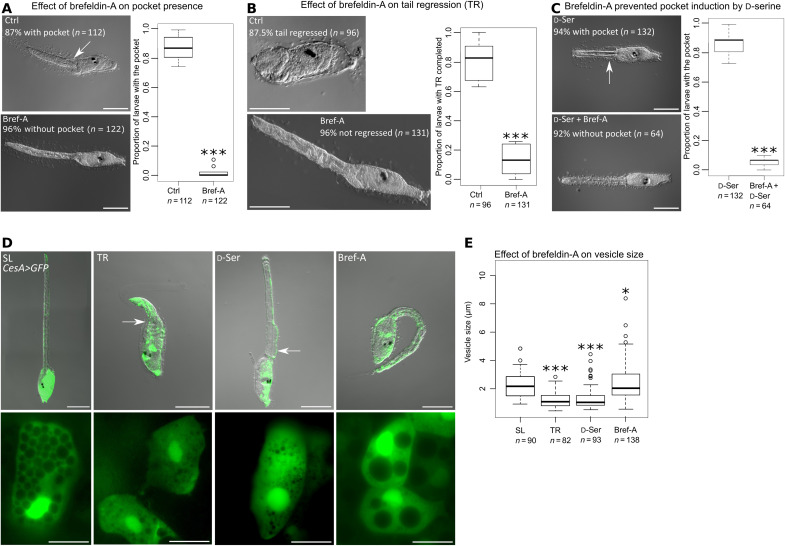
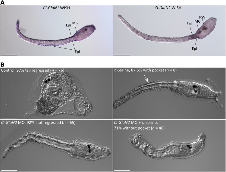
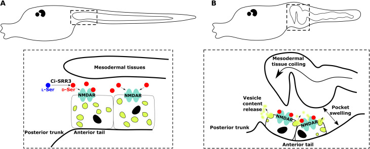
d-Serine, a free amino acid synthesized by serine racemase, is a coagonist of N-methyl-d-aspartate-type glutamate receptor (NMDAR). d-Serine in the mammalian central nervous system modulates glutamatergic transmission. Functions of d-serine in mammalian peripheral tissues such as skin have also been described. However, d-serine's functions in nonmammals are unclear. Here, we characterized d-serine-dependent vesicle release from the epidermis during metamorphosis of the tunicate Ciona. d-Serine leads to the formation of a pocket that facilitates the arrival of migrating tissue during tail regression. NMDAR is the receptor of d-serine in the formation of the epidermal pocket. The epidermal pocket is formed by the release of epidermal vesicles' content mediated by d-serine/NMDAR. This mechanism is similar to observations of keratinocyte vesicle exocytosis in mammalian skin. Our findings provide a better understanding of the maintenance of epidermal homeostasis in animals and contribute to further evolutionary perspectives of d-amino acid function among metazoans.


Figures

Fig. 1.
Fig. 1.. Characterization of Ci-SRR3 in Ciona larvae.
(A) The phylogenetic analysis of deuterostomian SRR and its close relative SDS, made by Bayesian inference. The SRR monophyletic group includes Ci-SRR1, Ci-SRR2, and Ci-SRR3. The topology was the same when Drosophila elegans SDS (XP_017118232.1) was used as the outgroup instead of switching D. melanogaster SRR (NP_731340.1). The alignment comprised 220 amino acids. The node robustness was determined by posterior probabilities. (B) Ciona SRR expression from hatched larvae [0 hours post-hatching (hph)] to larvae with tail regression in progress (Tail reg.) according to the RNA sequencing. The Ci-SRR3 expression significantly increases over time. (C) Expression of Ci-SRR3 relative to that of Ci-Ef1α as evaluated by quantitative reverse transcription polymerase chain reaction (RT-PCR). The expression of Ci-SRR3 significantly increased during the 12 hph. (D) Expression of Ci-SRR3 in a swimming larva, as revealed by whole-mount in situ hybridization (WISH). Ci-SRR3 is expressed in the epidermis (Epi) at the anterior tail and posterior trunk, in the endoderm (Endo), motor ganglion (MG), and posterior region of the sensory vesicle (PSV). Scale bar, 100 μm. (E) d-Serine concentration assay. The concentration of free d-serine remained stable during embryogenesis from the oocyte stage to the middle tailbud (MTB) and late tailbud (LTB) stages but significantly increased in 8 hph larvae. (F) Ci-SRR3 knockdown (KD) by the antisense morpholino oligonucleotide (MO) significantly decreased the quantity of d-serine in 8 hph larvae. The results of a cysteine assay used as the control did not reveal differences between the control larvae and the Ci-SRR3 MO-injected larvae. (G) The evaluation of Ci-SRR3 racemization activity, as revealed by the in vitro assay. Ci-SRR3 is capable of synthesizing d-serine from l-serine. (H) The relative expression level of Ci-SRR3 evaluated by the quantitative RT-PCR, comparing control larvae and Ci-gnrh2 KD larvae. The knockdown of Ci-gnrh2 significantly decreased the expression level of Ci-SRR3. *P < 0.05, **P < 0.01, ***P < 0.001.
Fig. 2.
Fig. 2.. Ci-SRR3 is necessary for pocket formation during metamorphosis.
(A) Control (Ctrl) and Ci-SRR3 MO-injected larvae during tail regression. Arrows indicate the pocket. (B) While tail regression (TR) was completed in the controls, a proportion of the MO-injected larvae had unabsorbed tail tip (arrow). The numbers of animals that completed the tail regression are shown in the panels. (C) A Ci-SRR3 KD juvenile exhibiting normal morphology except for the presence of the unabsorbed tail tip (arrow). (D) The proportion of larvae that completed the tail regression. The proportion was significantly lower in the MO-injected larvae compared with the controls. The addition of d-serine (d-ser) ameliorated the phenotype. (E) Wild-type larva during tail regression. The epidermis and inside tissues are separated by forming the pocket at the anterior region of the tail (arrows), a transparent space where migrating tissues coiled. Dotted lines indicate the junction between the tail and trunk. (F) Phalloidin labeling of larvae during tail regression. SRR3 MO-injected larvae did not have the pocket. The addition of d-serine led to a rescue of the pocket formation (arrows). (G) The proportion of larvae having a pocket was significantly reduced by the Ci-SRR3 MO. The phenotype was rescued by the addition of d-serine. Scale bars, 100 μm. *P < 0.05, **P < 0.01, ***P < 0.001.
Fig. 3.
Fig. 3.. d-Serine induces pocket formation without initiating metamorphosis.
(A) Treatments with d-serine (d-Ser), l-serine (l-Ser), and glycine (Gly) led to the formation of the pocket (arrows) in wild-type larvae that did not adhere to the substrate and therefore did not start metamorphosis. Glut, l-glutamate. (B) The effects of neurotransmitters on the induction of the pocket. (C) Phalloidin labeling exhibited the pocket induced by d-serine. The pocket can be anteriorly or posteriorly positioned. Most of the pockets induced by d-serine were positioned at the most anterior part of the tail. Scale bars, 200 μm (A) and 50 μm (C). *P < 0.05, **P < 0.01, ***P < 0.001.
Fig. 4.
Fig. 4.. The pocket is formed by the vesicle release from epidermal cells.
(A) Brefeldin-A (Bref-A) significantly prevented pocket (arrow) formation during tail regression (TR). (B) Brefeldin-A suppressed the completion of tail regression. (C) The formation of the pocket under d-serine treatment in nonsettled larvae was inhibited by brefeldin-A. (D) Visualization of epidermal vesicles in CesA>GFP electroporated larvae. Epidermal cells from the anterior tail of a swimming larva (SL) contained numerous large vesicles. During tail regression, epidermal cells surrounding the pocket (arrows) exhibited great reductions in vesicle size and number. When the pocket was induced by d-serine, the epidermal vesicles exhibited the same characteristics as the larvae during tail regression. Brefeldin-A–treated larvae presented large epidermal vesicles. (E) Measurement of the epidermal vesicle size. Compared with the swimming larvae, the larvae with tail regression in progress and under d-serine treatment had smaller vesicles. Conversely, the brefeldin-A–treated larvae exhibited larger vesicles. Scale bars, 100 μm (A to D, top) and 20 μm (D, bottom). *P < 0.05, **P < 0.01, ***P < 0.001.
Fig. 5.
Fig. 5.. NMDAR is the receptor of d-serine for epidermal pocket formation.
(A) The expression patterns of Ci-GluN1 and Ci-GluN2 at the larval stage, as revealed by WISH. Ci-GluN1 is expressed in the tail epidermis (Epi) and motor ganglion (MG). Ci-GluN2 is expressed in the anterior tail epidermis, motor ganglion, and posterior region of the sensory vesicle (PSV). (B) Ci-GluN2 MO blocked tail regression and prevented pocket formation. d-Serine failed to induce the pocket in Ci-GluN2 MO-injected larvae. Scale bars, 100 μm.
Fig. 6.
Fig. 6.. Mechanisms of the epidermal pocket formation during Ciona tail regression.
(A) d-Serine is synthetized by Ci-SRR3 from l-serine. At the beginning of tail regression, the NMDA receptors from epidermal cells at the most anterior part of the tail are activated by d-serine. (B) During tail regression, activation of NMDA receptors led to the release of the epidermal cell vesicle content toward the extracellular matrix. Accumulation of vesicle content intro extracellular matrix allows separation between epidermis and inside tissues. Concomitantly, epidermal tissues swell, creating an empty space for welcoming of migrating tissues that start to coil. In dotted squares, epidermal cells are intentionally not to scale to show in-depth mechanism.

Similar articles

Cited by

References

    1. Hashimoto A., Nishikawa T., Hayashi T., Fujii N., Harada K., Oka T., Takahashi K., The presence of free D-serine in rat brain. FEBS Lett. 296, 33–36 (1992). - PubMed
    1. Wolosker H., Sheth K. N., Takahashi M., Mothet J. P., Brady R. O., Ferris C. D., Snyder S. H., Purification of serine racemase: Biosynthesis of the neuromodulator D-serine. Proc. Natl. Acad. Sci. U.S.A. 96, 721–725 (1999). - PMC - PubMed
    1. Mothet J. P., Parent A. T., Wolosker H., Brady R. O., Linden D. J., Ferris C. D., Rogawski M. A., Snyder S. H., D-serine is an endogenous ligand for the glycine site of the N-methyl-D-aspartate receptor. Proc. Natl. Acad. Sci. U.S.A. 97, 4926–4931 (2000). - PMC - PubMed
    1. Matsui T., Sekiguchi M., Hashimoto A., Tomita U., Nishikawa T., Wada K., Functional comparison of D-serine and glycine in rodents: The effect on cloned NMDA receptors and the extracellular concentration. J. Neurochem. 65, 454–458 (1995). - PubMed
    1. Priestley T., Laughton P., Myers J., Le Bourdellés B., Kerby J., Whiting P. J., Pharmacological properties of recombinant human N-methyl-D-aspartate receptors comprising NR1a/NR2A and NR1a/NR2B subunit assemblies expressed in permanently transfected mouse fibroblast cells. Mol. Pharmacol. 48, 841–848 (1995). - PubMed

Publication types