. 2022 Jan 18;14(3):424.
doi: 10.3390/nu14030424.

Co-toxicity of Endotoxin and Indoxyl Sulfate, Gut-Derived Bacterial Metabolites, to Vascular Endothelial Cells in Coronary Arterial Disease Accompanied by Gut Dysbiosis

Affiliations

Co-toxicity of Endotoxin and Indoxyl Sulfate, Gut-Derived Bacterial Metabolites, to Vascular Endothelial Cells in Coronary Arterial Disease Accompanied by Gut Dysbiosis

Marcin Choroszy et al. Nutrients. .

Abstract

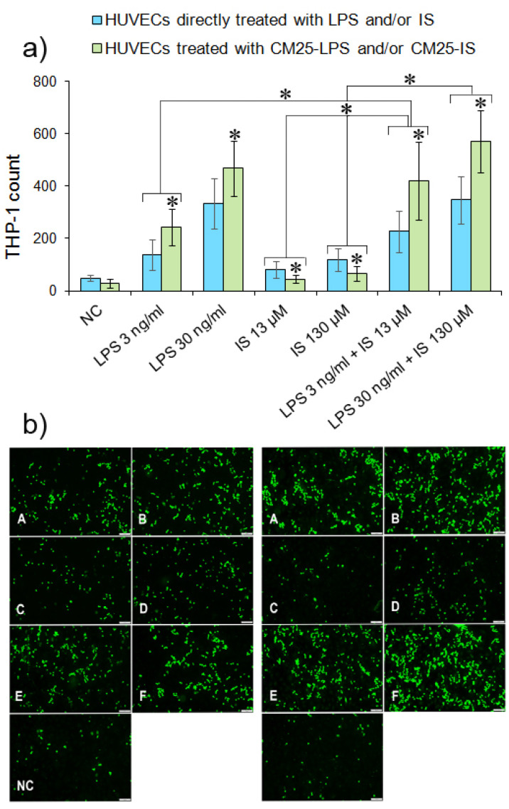
Gut dysbiosis, alongside a high-fat diet and cigarette smoking, is considered one of the factors promoting coronary arterial disease (CAD) development. The present study aimed to research whether gut dysbiosis can increase bacterial metabolites concentration in the blood of CAD patients and what impact these metabolites can exert on endothelial cells. The gut microbiomes of 15 age-matched CAD patients and healthy controls were analyzed by 16S rRNA sequencing analysis. The in vitro impact of LPS and indoxyl sulfate at concentrations present in patients' sera on endothelial cells was investigated. 16S rRNA sequencing analysis revealed gut dysbiosis in CAD patients, further confirmed by elevated LPS and indoxyl sulfate levels in patients' sera. CAD was associated with depletion of Bacteroidetes and Alistipes. LPS and indoxyl sulfate demonstrated co-toxicity to endothelial cells inducing reactive oxygen species, E-selectin, and monocyte chemoattractant protein-1 (MCP-1) production. Moreover, both of these metabolites promoted thrombogenicity of endothelial cells confirmed by monocyte adherence. The co-toxicity of LPS and indoxyl sulfate was associated with harmful effects on endothelial cells, strongly suggesting that gut dysbiosis-associated increased intestinal permeability can initiate or promote endothelial inflammation and atherosclerosis progression.

Keywords: Bacteroidetes; LPS; coronary artery disease; dysbiosis; gut microbiome; indoxyl sulfate; obesity.


Conflict of interest statement

The authors declare no known competing financial interests or personal relationships that could influence the work reported in this paper.

Figures

Figure 1
Figure 1
Community abundance (%) in gut microbiome of CAD patients and healthy controls (HC) at the phylum level. The asterisk indicates a statistically significant difference (p < 0.05) between CAD and HC groups.
Figure 2
Figure 2
The most notable differences at the phylum, class, order, family and genus levels in the gut microbiome of CAD patients compared to healthy controls. The asterisks above bars indicate statistically significant differences (p < 0.05) between CAD and HC groups.
Figure 3
Figure 3
Enterotypes (%) distribution in CAD and HC groups. Enterotype I—Bacteroides prevalence; enterotype II—prevotella prevalence, and enterotype III Ruminococcus prevalence.
Figure 4
Figure 4
HUVECs and monocyte-derived macrophages (MDM) viability after stimulation with LPS and indoxyl sulfate (IS). (a) HUVECs and (b) MDM viability was assessed after 18 h with MTT. Data are means ± SEM of three independent experiments performed in triplicate. The asterisk above the bar indicates a statistically significant difference (p < 0.05).
Figure 5
Figure 5
HUVECs morphology after stimulation with LPS and indoxyl. HUVECs were treated with LPS and IS or their combinations (a) or with conditioned medium from MDM (b) for 18 h. Fluorescence images present cells treated with LPS at 3 ng/mL and 30 ng/mL (Panels A,B); cells treated with indoxyl at 13 µM and 130 µM (Panels C,D); cells treated with LPS 3 ng/mL + IS 13 µM and LPS30 ng/mL + IS 130 µM (Panels E,F); and untreated cells (Panels G). Cells were stained with phalloidin-FITC and DAPI to visualize the cell’s actin filaments and nuclei, respectively.
Figure 6
Figure 6
Reactive oxygen species (ROS) production. (a) HUVECs were treated for 5 h with LPS or IS or combined metabolites at appropriate concentrations. ROS was evaluated in HUVECs using a permeable probe H2DCF-DA compared to untreated cells (NC) as a negative control. Fluorescence intensity was read spectrophotometrically at Ex/EM = 488/525 nm. Data are presented as means ± SEM from three independent assays performed in triplicate. The asterisks above bars indicate statistically significant differences between negative control and metabolites examined. (b) ROS production was visualized in a fluorescent microscope. The image shows HUVECs treated with metabolites at the following concentrations: LPS 3 ng/mL and 30 ng/mL (panels A,B); IS 13 µM and 130 µM (panels C,D); LPS 3 ng/mL + IS 13 µM and LPS 30 ng/mL + IS 130 µM (panels E,F); and negative control (panel G).
Figure 7
Figure 7
E-selectin and MCP-1 production in HUVECs stimulated with LPS and indoxyl. (a) E-selectin levels in HUVECs treated directly (blue bars) with LPS and IS or both metabolites and indirectly (green bars) with CM25-LPS/CM25-IS at appropriate concentrations. The asterisks above bars indicate the statistical differences in E-selectin levels between HUVECs stimulated directly with LPS/IS or indirectly with CM25. E-selectin was evaluated in HUVECs stimulated with LPS or IS or both bacterial metabolites for 18 h with FITC-conjugated mouse monoclonal anti-human E-selectin antibody. Data are presented as means ± SEM from two independent assays performed in quadruplicate. (b) MPC-1 levels in CM25 from MDM (black bars) and a culture media from HUVECs stimulated for 18 h with CM25 (green bars) or with LPS/IS (blue bars). The asterisks above black bars represent statistically significant differences between MCP-1 levels in CM25 and a culture media from HUVECs stimulated with LPS and IS for 18 h. The asterisks above white and grey bars indicate differences between MCP-1 levels in a culture media from HUVECs stimulated with CM25 and LPS/IS. MCP-1 was evaluated using the MCP-1 Human ELISA kit. Data are the means ± SEM from two independent assays performed in duplicate.
Figure 8
Figure 8
Monocyte adherence to HUVECs stimulated with LPS and indoxyl. (a) Quantitative THP-1 adherence to HUVECs treated directly (blue bars) with LPS/IS or indirectly (green bars) with CM25-LPS/CM25-IS at appropriate concentrations. The asterisks above bars indicate statistically significant differences between HUVECs treated directly and indirectly. The asterisks above white and grey bars indicate statistically significant differences between results for directly and indirectly treated cells. THP-1 cells stained with calcein-AM were incubated with HUVECs for 4 h and counted in a fluorescent microscope after washing out. (b) Fluorescence images of calcein-AM-stained monocytes adhering to HUVECs present HUVECs pretreated with LPS at 3 ng/mL and 30 ng/mL (panels A,B); cells treated with indoxyl at 13 µM and 130 µM (Panels C,D); cells treated with LPS 3 ng/mL + IS 13 µM and LPS30 ng/mL + IS 130 µM (Panels E,F); and untreated cells (NC).

References

    1. Vasan R.S., Benjamin E.J. The Future of Cardiovascular Epidemiology. Circulation. 2016;133:2626–2633. doi: 10.1161/CIRCULATIONAHA.116.023528. - DOI - PMC - PubMed
    1. Trøseid M., Andersen G.Ø., Broch K., Hov J.R. The gut microbiome in coronary artery disease and heart failure: Current knowledge and future directions. EBioMedicine. 2020;52:102649. doi: 10.1016/j.ebiom.2020.102649. - DOI - PMC - PubMed
    1. Tang W.H., Kitai T., Hazen S.L. Gut microbiota in cardiovascular health and disease. Circulation Res. 2017;120:1183–1196. doi: 10.1161/CIRCRESAHA.117.309715. - DOI - PMC - PubMed
    1. Lobionda S., Sittipo P., Kwon H.Y., Lee Y.K. The role of gut microbiota in intestinal inflammation with respect to diet and extrinsic stressors. Microorganisms. 2019;7:271. doi: 10.3390/microorganisms7080271. - DOI - PMC - PubMed
    1. Fujii H., Goto S., Fukagawa M. Role of uremic toxins for kidney, cardiovascular, and bone dysfunction. Toxins. 2018;10:202. doi: 10.3390/toxins10050202. - DOI - PMC - PubMed