. 2022 Jan 29;14(3):599.
doi: 10.3390/nu14030599.

One-Carbon Metabolism in Alzheimer's Disease and Parkinson's Disease Brain Tissue

Affiliations

One-Carbon Metabolism in Alzheimer's Disease and Parkinson's Disease Brain Tissue

Karel Kalecký et al. Nutrients. .

Abstract

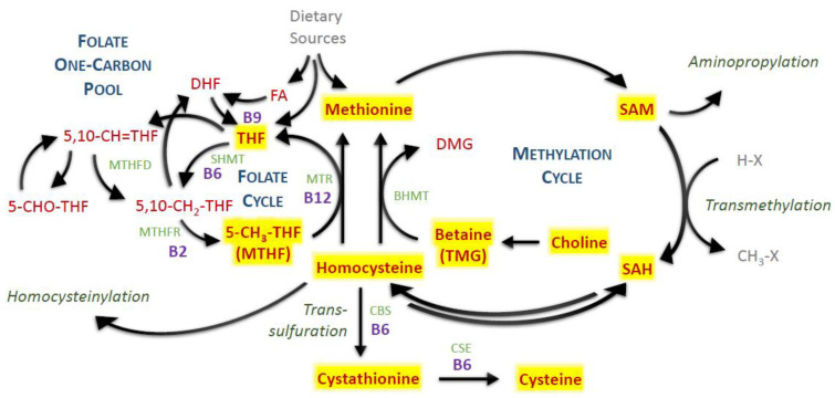
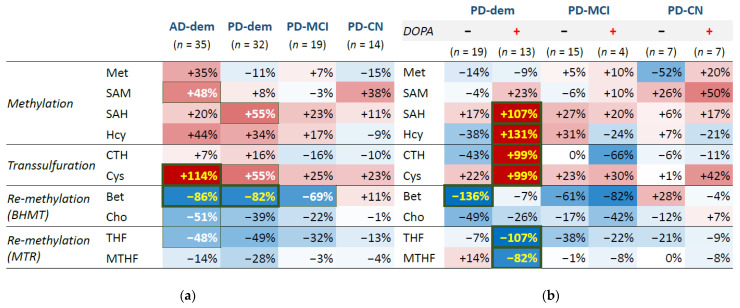
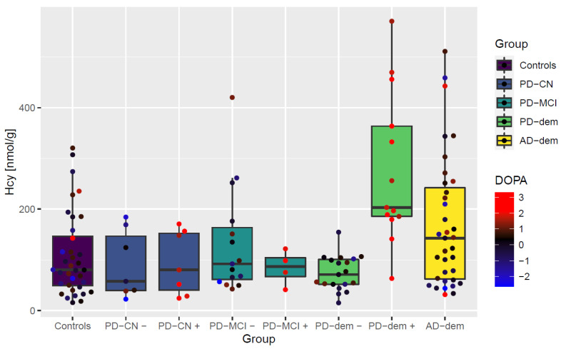
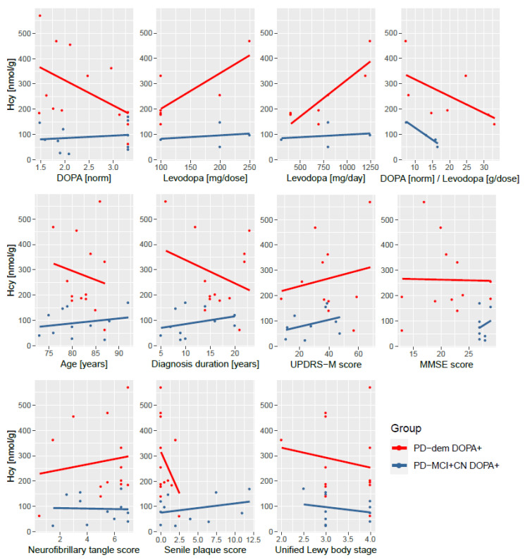
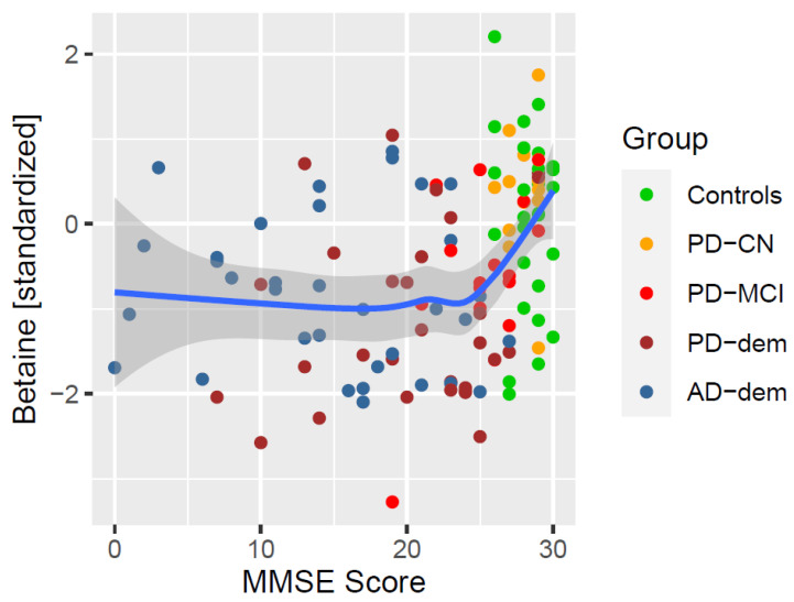
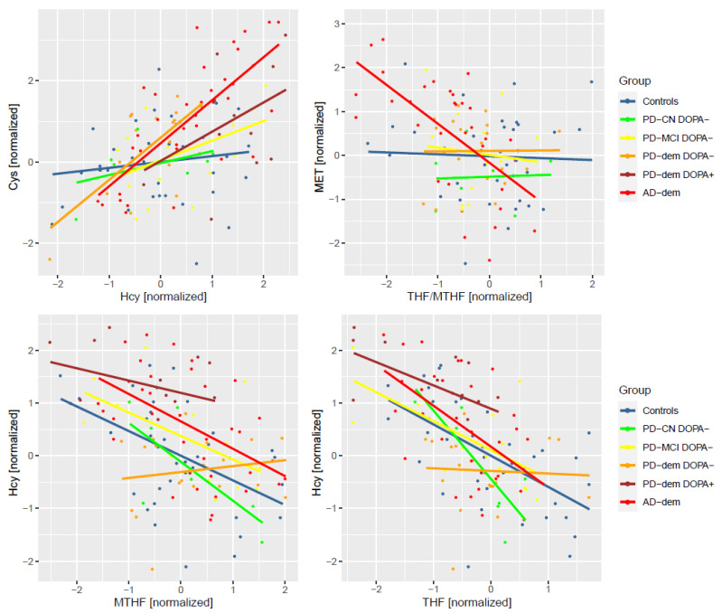
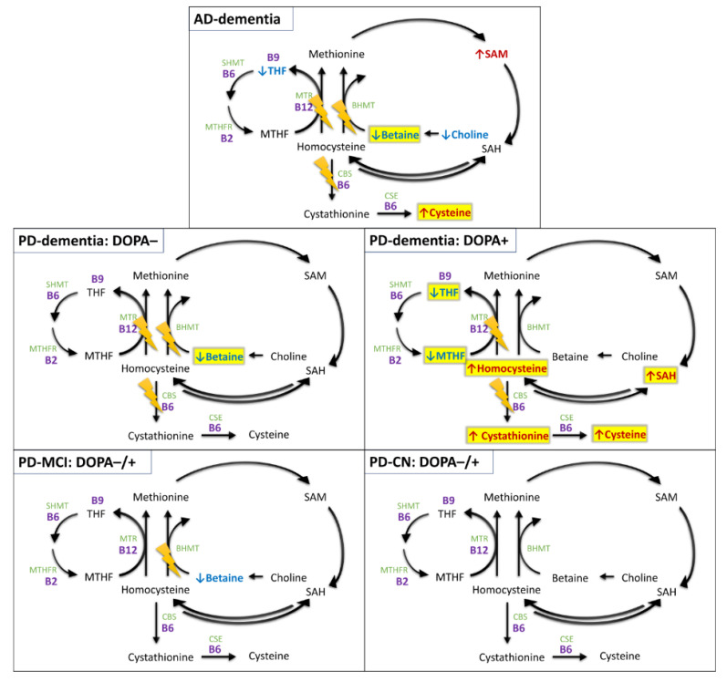
Disruptions in one-carbon metabolism and elevated homocysteine have been previously implicated in the development of dementia associated with Alzheimer's disease (AD) and Parkinson's disease (PD). Moreover, a PD diagnosis itself carries substantial risk for the development of dementia. This is the first study that explores alterations in one-carbon metabolism in AD and PD directly in the human brain frontal cortex, the primary center of cognition. Applying targeted liquid chromatography-tandem mass spectrometry (LC-MS/MS), we analyzed post-mortem samples obtained from 136 subjects (35 AD, 65 PD, 36 controls). We found changes in one-carbon metabolites that indicate inefficient activation of cystathionine β-synthase (CBS) in AD and PD subjects with dementia, the latter seemingly accompanied by a restricted re-methylation flow. Levodopa-carbidopa is known to reduce available vitamin B6, which would explain the hindered CBS activity. We present evidence of temporary non-protein-bound homocysteine accumulation upon levodopa intake in the brain of PD subjects with dementia but not in non-demented PD subjects. Importantly, this homocysteine elevation is not related to levodopa dosage, disease progression, or histopathological markers but exclusively to the dementia status. We hypothesize that this levodopa-induced effect is a direct cause of dementia in PD in susceptible subjects with reduced re-methylation capacity. Furthermore, we show that betaine best correlates with cognitive score even among PD subjects alone and discuss nutritional recommendations to improve one-carbon metabolism function.

Keywords: Alzheimer’s disease; Parkinson’s disease; betaine; brain frontal cortex; dementia; homocysteine; levodopa; metabolomics; one-carbon metabolism.


Conflict of interest statement

The authors declare no conflict of interest. The funders had no role in the design of the study; in the collection, analyses, or interpretation of data; in the writing of the manuscript; or in the decision to publish the results.

Figures

Figure 1
Figure 1
One-carbon metabolism. Red—measured metabolites; yellow highlighted—detected and quantified metabolites; light green—enzymes; purple—vitamin B cofactors. Abbreviations: BHMT, betaine-homocysteine methyltransferase; CBS, cystathionine β-synthase; CSE, cystathionine γ-lyase; DHF, dihydrofolate; DMG, dimethylglycine; FA, folic acid; MTHF, methyltetrahydrofolate; MTHFD, methylenetetrahydrofolate dehydrogenase; MTHFR, methylenetetrahydrofolate reductase; MTR, methionine synthase; SAH, S-adenosylhomocysteine; SAM, S-adenosylmethionine; SHMT, serine hydroxymethyltransferase; THF, tetrahydrofolate; TMG, trimethylglycine; X, methylation acceptor residue.
Figure 2
Figure 2
Group heatmap for differential analysis by diagnosis groups. Regardless of current levodopa status (a) and with levodopa interaction (b) as identified from DOPA concentrations. Red—upregulation; blue—downregulation. Font color: yellow—FDR ≤ 0.05; white—p-value ≤ 0.05; black—p-value > 0.05.
Figure 3
Figure 3
Boxplot of non-protein-bound homocysteine levels by diagnostic group with levodopa interaction. Points represent individual subjects. + and − signs attached as group suffixes denote DOPA+ and DOPA− subgroups. The measured DOPA levels (transformed and standardized) are further visualized as the point color.
Figure 4
Figure 4
Independence of non-protein-bound homocysteine elevation upon levodopa intake in PD subjects with dementia on medication dosage and measures of disease progression. (Top): measured DOPA, levodopa medication amount per dose and per day and a ratio of measured DOPA and levodopa dosage; (middle row): age, length of the diagnosis, UPDRS-M motor score, and MMSE cognitive score; (bottom): histopathological scores of neurofibrillary tangle density, senile plaque density, and Unified Lewy body stage. The DOPA+ PD subjects with dementia (red) have increased homocysteine levels than non-demented DOPA+ PD subjects (blue) irrespective of these variables. Levodopa dosage, UPDRS-M and MMSE scores are not available for all subjects. Abbreviations: norm, normalized (transformed and standardized).
Figure 5
Figure 5
Association of MMSE with betaine. The blue line represents a locally estimated scatterplot smoothing (LOESS) trend with 95% confidence interval shown as the dark gray area.
Figure 6
Figure 6
Differentially altered metabolic correlations by diagnosis group. Only DOPA− subjects are included to avoid levodopa-related pathway disturbances. PD-dem DOPA+ subjects are included for comparison with the effects presented for DOPA− PD subjects with dementia to demonstrate their similarity (apart from elevated Hcy) even upon levodopa intake.
Figure 7
Figure 7
Overview of differentially expressed metabolites and detected pathway abnormalities by group and levodopa status. Upregulated (red) and downregulated (blue) metabolites with p-value ≤ 0.05; additionally highlighted with yellow for FDR ≤ 0.05. The lightning icons denote potentially impacted pathways (regardless of primary causality) based on abnormalities found in differential analysis and correlation analysis. Notice that the alterations revealed in DOPA− PD subjects with dementia are well reflected in metabolite concentrations after levodopa-induced Hcy challenge. Light green—enzymes; purple—vitamin B cofactors. Abbreviations: BHMT, betaine-homocysteine methyltransferase; CBS, cystathionine β-synthase; CSE, cystathionine γ-lyase; MTHF, methyltetrahydrofolate; MTHFR, methylenetetrahydrofolate reductase; MTR, methionine synthase; SAH, S-adenosylhomocysteine; SAM, S-adenosylmethionine; SHMT, serine hydroxymethyltransferase; THF, tetrahydrofolate.

References

    1. Perła-Kaján J., Twardowski T., Jakubowski H. Mechanisms of homocysteine toxicity in humans. Amino Acids. 2007;32:561–572. doi: 10.1007/s00726-006-0432-9. - DOI - PubMed
    1. Zieminska E., Lazarewicz J.W. Excitotoxic neuronal injury in chronic homocysteine neurotoxicity studied in vitro: The role of NMDA and group I metabotropic glutamate receptors. Acta Neurobiol. Exp. Wars. 2006;66:301–309. - PubMed
    1. Cacciari E., Salardi S. Clinical and laboratory features of homocystinuria. Haemostasis. 1989;19((Suppl. 1)):10–13. doi: 10.1159/000216090. - DOI - PubMed
    1. Graham I.M., Daly L.E., Refsum H.M., Robinson K., Brattström L.E., Ueland P.M., Palma-Reis R.J., Boers G.H., Sheahan R.G., Israelsson B., et al. Plasma homocysteine as a risk factor for vascular disease. The European Concerted Action Project. JAMA. 1997;277:1775–1781. doi: 10.1001/jama.1997.03540460039030. - DOI - PubMed
    1. Virtanen J.K., Voutilainen S., Happonen P., Alfthan G., Kaikkonen J., Mursu J., Rissanen T.H., Kaplan G.A., Korhonen M.J., Sivenius J., et al. Serum homocysteine, folate and risk of stroke: Kuopio Ischaemic Heart Disease Risk Factor (KIHD) Study. Eur. J. Cardiovasc. Prev. Rehabil. 2005;12:369–375. doi: 10.1097/01.hjr.0000160834.75466.b0. - DOI - PubMed