

Parvovirus enteritis and other risk factors associated with persistent gastrointestinal signs in dogs later in life: a retrospective cohort study
- PMID: 35277172
- PMCID: PMC8915519
- DOI: 10.1186/s12917-022-03187-7
Parvovirus enteritis and other risk factors associated with persistent gastrointestinal signs in dogs later in life: a retrospective cohort study
Abstract
Background: Parvoviral enteritis (PE) is a viral gastrointestinal (GI) infection of dogs. Recovery from PE has been associated with persistent GI signs later in life. The objectives of this study were: (i) To determine whether dogs that have recovered from PE (post-parvo dogs) had an increased risk of persistent GI signs compared to uninfected control dogs. (ii) To investigate the lifestyle and clinicopathologic factors that are associated with persistent GI signs in post-parvo dogs.
Methods: A total of 86 post-parvo dogs and 52 age-matched control dogs were enrolled in this retrospective cohort study. Many years after hospitalization for PE, the owners were interviewed about the health and habits of their dogs using a questionnaire. We used generalized linear mixed effects models to test whether parvovirus enteritis and other risk factors are associated with owner-recognized general health problems in all dogs and with owner-recognized persistent GI signs in post-parvo dogs.
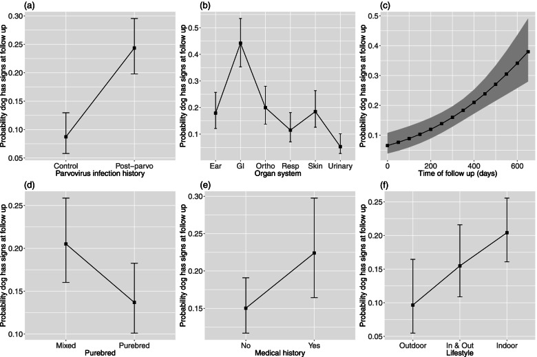
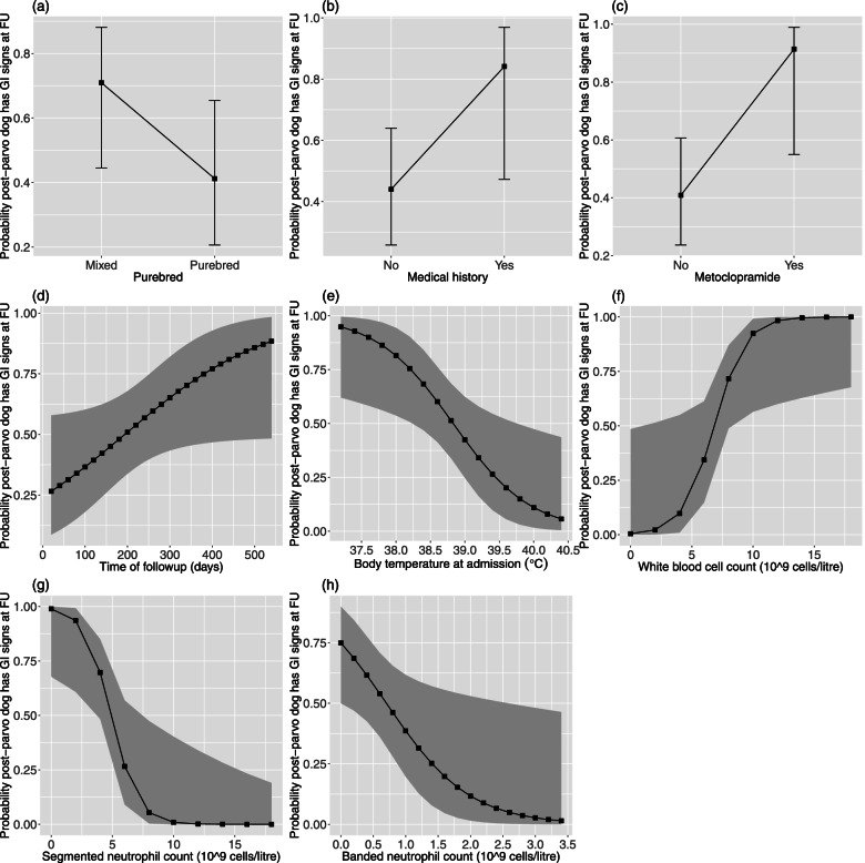
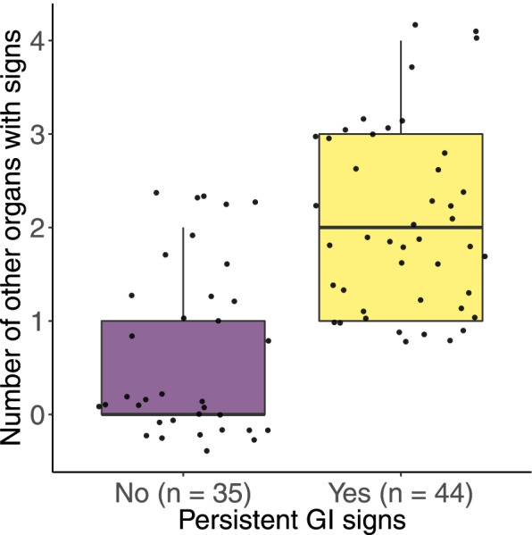
Results: The prevalence of persistent GI signs was significantly higher in post-parvo dogs compared to control dogs (57% vs 25%, P < 0.001). Markers of disease severity at the time of hospital admission such as neutropenia, low body temperature (BT), and treatment with an antiemetic medication (metoclopramide) were significant risk factors for persistent GI signs in post-parvo dogs. For example, PE-affected dogs that were hypothermic at hospital admission (BT of 37.2 °C) were 16.6 × more likely to have GI signs later in life compared to hyperthermic dogs (BT of 40.4 °C). The presence of persistent GI signs in post-parvo dogs was a risk factor for health problems in other organ systems.
Conclusions: Parvovirus enteritis is a significant risk factor for persistent GI signs in dogs highlighting the importance of prevention. The risk factors identified in the present study may guide future investigations on the mechanisms that link parvovirus enteritis to chronic health problems in dogs.
Keywords: Antimicrobials; Canine; Diarrhea; Gastrointestinal system; Immunology; Metoclopramide; Parvovirus; Vomiting.
© 2022. The Author(s).
Conflict of interest statement
The authors declare that they have no competing interests.
Figures
Similar articles
-
Characterization of the use of antiemetic agents in dogs with parvoviral enteritis treated at a veterinary teaching hospital: 77 cases (1997-2000).J Am Vet Med Assoc. 2005 Dec 1;227(11):1787-93. doi: 10.2460/javma.2005.227.1787. J Am Vet Med Assoc. 2005. PMID: 16342528
-
Amelioration of oxidative stress using N-acetylcysteine in canine parvoviral enteritis.J Vet Pharmacol Ther. 2018 Feb;41(1):68-75. doi: 10.1111/jvp.12434. Epub 2017 Jul 12. J Vet Pharmacol Ther. 2018. PMID: 28703421 Free PMC article. Clinical Trial.
-
Canine parvovirus.Vet Clin North Am Small Anim Pract. 2010 Nov;40(6):1041-53. doi: 10.1016/j.cvsm.2010.07.007. Vet Clin North Am Small Anim Pract. 2010. PMID: 20933134 Review.
-
Recombinant bactericidal/permeability-increasing protein (rBPI21) for treatment of parvovirus enteritis: a randomized, double-blinded, placebo-controlled trial.J Vet Intern Med. 2001 Jul-Aug;15(4):355-60. J Vet Intern Med. 2001. PMID: 11467593 Clinical Trial.
-
Update on Canine Parvoviral Enteritis.Vet Clin North Am Small Anim Pract. 2025 May;55(3):405-425. doi: 10.1016/j.cvsm.2025.01.002. Epub 2025 Mar 4. Vet Clin North Am Small Anim Pract. 2025. PMID: 40044515 Review.
Cited by
-
Prevalence and significance of a canine bocavirus-2 outbreak in a cohort of military dogs in Austria.Front Vet Sci. 2024 Sep 5;11:1461136. doi: 10.3389/fvets.2024.1461136. eCollection 2024. Front Vet Sci. 2024. PMID: 39301279 Free PMC article.
-
Long-Term Follow-Up After Acute Gastroenteritis Caused by Giardia Infection in Juvenile Dogs.J Vet Intern Med. 2025 Jul-Aug;39(4):e70123. doi: 10.1111/jvim.70123. J Vet Intern Med. 2025. PMID: 40448678 Free PMC article.
-
Environmental risk factors in puppies and kittens for developing chronic disorders in adulthood: A call for research on developmental programming.Front Vet Sci. 2022 Dec 23;9:944821. doi: 10.3389/fvets.2022.944821. eCollection 2022. Front Vet Sci. 2022. PMID: 36619947 Free PMC article. Review.
-
Prognostic factors associated with survival and hospitalization time in pediatric canine patients diagnosed with presumptive acute viral gastroenteritis.Vet World. 2022 Aug;15(8):2095-2101. doi: 10.14202/vetworld.2022.2095-2101. Epub 2022 Aug 30. Vet World. 2022. PMID: 36313832 Free PMC article.
References
MeSH terms
LinkOut - more resources
Full Text Sources
