Review
. 2022 Mar 11;41(1):92.
doi: 10.1186/s13046-022-02281-w.

Recent advances in the developmental origin of neuroblastoma: an overview

Affiliations
Review

Recent advances in the developmental origin of neuroblastoma: an overview

Mirco Ponzoni et al. J Exp Clin Cancer Res. .

Abstract

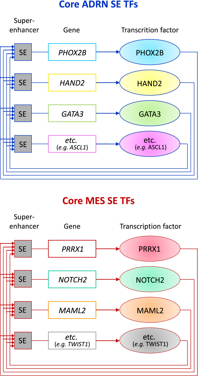
Neuroblastoma (NB) is a pediatric tumor that originates from neural crest-derived cells undergoing a defective differentiation due to genomic and epigenetic impairments. Therefore, NB may arise at any final site reached by migrating neural crest cells (NCCs) and their progeny, preferentially in the adrenal medulla or in the para-spinal ganglia.NB shows a remarkable genetic heterogeneity including several chromosome/gene alterations and deregulated expression of key oncogenes that drive tumor initiation and promote disease progression.NB substantially contributes to childhood cancer mortality, with a survival rate of only 40% for high-risk patients suffering chemo-resistant relapse. Hence, NB remains a challenge in pediatric oncology and the need of designing new therapies targeted to specific genetic/epigenetic alterations become imperative to improve the outcome of high-risk NB patients with refractory disease or chemo-resistant relapse.In this review, we give a broad overview of the latest advances that have unraveled the developmental origin of NB and its complex epigenetic landscape.Single-cell RNA sequencing with spatial transcriptomics and lineage tracing have identified the NCC progeny involved in normal development and in NB oncogenesis, revealing that adrenal NB cells transcriptionally resemble immature neuroblasts or their closest progenitors. The comparison of adrenal NB cells from patients classified into risk subgroups with normal sympatho-adrenal cells has highlighted that tumor phenotype severity correlates with neuroblast differentiation grade.Transcriptional profiling of NB tumors has identified two cell identities that represent divergent differentiation states, i.e. undifferentiated mesenchymal (MES) and committed adrenergic (ADRN), able to interconvert by epigenetic reprogramming and to confer intra-tumoral heterogeneity and high plasticity to NB.Chromatin immunoprecipitation sequencing has disclosed the existence of two super-enhancers and their associated transcription factor networks underlying MES and ADRN identities and controlling NB gene expression programs.The discovery of NB-specific regulatory circuitries driving oncogenic transformation and maintaining the malignant state opens new perspectives on the design of innovative therapies targeted to the genetic and epigenetic determinants of NB. Remodeling the disrupted regulatory networks from a dysregulated expression, which blocks differentiation and enhances proliferation, toward a controlled expression that prompts the most differentiated state may represent a promising therapeutic strategy for NB.

Keywords: Adrenergic; Core regulatory circuitries; Mesenchymal; Neural crest; Neuroblastoma; Transcription factors.


Conflict of interest statement

The authors declare no competing interests.

Figures

Fig. 1
Fig. 1
Early stages of physiologic development of NC-derived lineages. Summary of the main processes and gene pathways involved in the early steps of physiologic development of NC-derived lineages emerging from mouse studies and common in mammals. Specifier transcription factors are reported in boxes in hierarchical order. Illustrations has been retrieved from https://app.biorender.com
Fig. 2
Fig. 2
Cell fates and multistep differentiation processes in physiologic development of the sympatho-adrenal lineage in mice. Cell populations and transitions, here illustrated, are based on scRNA-seq studies with spatial transcriptomics and lineage tracing conducted in developing mouse embryos. In the developing AM there is a co-localization of sympathetic and SCP-derived components at different degrees of differentiation, outlined as solid painted circles. Cell type populations involved in physiologic development of the sympatho-adrenal lineage include cell progenitors, transient cells, and cells at different developmental stages toward the final differentiated cell types. Embryonic developmental mouse timeline expressed in days: E10.5, E11.5, E13.5. Basic cell icons have been retrieved from https://app.biorender.com and modified by Adobe Photoshop CC 2019
Fig. 3
Fig. 3
Cell fates and multistep differentiation processes in physiologic development of the sympatho-adrenal lineage in humans. Cell type populations involved in physiologic development of the sympatho-adrenal lineage, here illustrated, include: bipotent cell progenitors, sympatho-adrenal (SA) progenitors, and SNPCs, as emerged from lineage tracing in mouse embryos; SCPs, transient cells and cells at different developmental stages toward the final differentiated state, based on scRNA-seq studies with spatial transcriptomics conducted in human embryos. In the developing adrenal medulla (AM) there is a co-localization of SCP-derived components, sympathetic and chromaffin at different degrees of differentiation, which are outlined as solid painted circles. Developmental human timeline expressed as wpc: weeks post conception. Basic cell icons have been retrieved from https://app.biorender.com and next modified by Adobe Photoshop CC 2019
Fig. 4
Fig. 4
Lineage-specific CRC and SE-associated TFs in MES and ADRN NB cells. Lineage-specific CRCs of ADRN and MES cell identities in NB are composed of a set of SE-associated lineage TFs that bind to each other and induce a powerful feed-forward loop. Illustration modified from Tim J. B. van Groningen, 2020 [94]
Fig. 5
Fig. 5
Enhancer/super-enhancer-driven transcription of ADRN CRC genes by MYCN invasion. A Schematic representation of the ADRN CRC composed of a set of super-enhancer (SE)-associated lineage TFs (e.g. PHOX2B, GATA3, HAND2, etc.) invaded by MYCN (red arrows) following MYCN amplification/overexpression. Each TF (ovals) binds its own SE and each of the other SEs belonging to the CRC, thus inducing a powerful feed-forward loop in all the other genes. B Structural drawing of the molecular interactions within the transcriptional unit of a gene belonging to the ADNR CRC (cyan box). The white circle indicates a general TF bound to the promoter site to activate transcription. Starting from the 5′ end, an SE of the ADRN CRC (as indicated in the upper part A) consists of a clustering of E-boxes in close genomic proximity to each other (e.g. three in this figure) bound by all TFs taking part in the ADRN CRC (soft color circles) and invaded by MYCN (red solid circles) when it is amplified/overexpressed. Thanks to the recruitment of the mediator (Med) complex, an SE can interact with the basal transcription machinery and RNA polymerase II (Pol II) at the promoter of the target gene through a looping process to drive its transcription. In addition, overexpressed MYCN can bind a low-affinity E-box or an enhancer of the target gene, thus strongly activating its transcription (thick arrow) and increasing the amount of the gene products (cyan solid circles). The enhancer can be co-occupied by additional TFs together with MYCN (e.g. HAND2 or TWIST1, yellow-orange circles). Invasion of regulatory elements by increased levels of MYCN lead to an enhancer/SE-driven transcription of the target gene. TFs that activate transcription are indicated with “+” within circles. Illustration slightly modified from Perri et al., 2021 [5]
Fig. 6
Fig. 6
Scheme of the cell composition in human developing AM and in NB risk subtypes. Physiologic developmental cell composition of AM during consecutive differentiation stages (upper part, blue arrows). Genetic alterations or epigenetic perturbations at different stages of AM differentiation may hit different cell types promoting cell selection and malignant transformation (red arrows, lower part). Illustration modified from Hermann Rohrer, 2021 [86]

References

    1. Padovan-Merhar OM, Raman P, Ostrovnaya I, Kalletla K, Rubnitz KR, Sanford EM, et al. Enrichment of targetable mutations in the relapsed neuroblastoma genome. Plos Genet. 2016;12(12):e1006501. - PMC - PubMed
    1. Maris JM, Hogarty MD, Bagatell R, Cohn SL. Neuroblastoma. Lancet. 2007;369(9579):2106–2120. - PubMed
    1. Matthay KK, Maris JM, Schleiermacher G, Nakagawara A, Mackall CL, Diller L, et al. Neuroblastoma. Nat Rev Dis Primers. 2016;2:16078. - PubMed
    1. Maris JM. Recent advances in neuroblastoma. N Engl J Med. 2010;362(23):2202–2211. - PMC - PubMed
    1. Perri P, Ponzoni M, Corrias MV, Ceccherini I, Candiani S, Bachetti T. A focus on regulatory networks linking micrornas, transcription factors and target genes in neuroblastoma. Cancers (Basel). 2021;13(21):5528. - PMC - PubMed

Substances