. 2022 Mar 11;14(1):15.
doi: 10.1038/s41368-022-00165-5.

Microenvironmental stiffness mediates cytoskeleton re-organization in chondrocytes through laminin-FAK mechanotransduction

Affiliations

Microenvironmental stiffness mediates cytoskeleton re-organization in chondrocytes through laminin-FAK mechanotransduction

Chenchen Zhou et al. Int J Oral Sci. .

Abstract

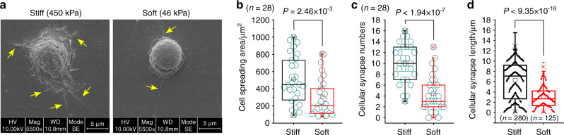
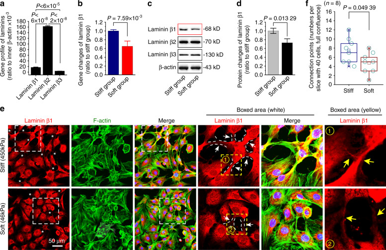
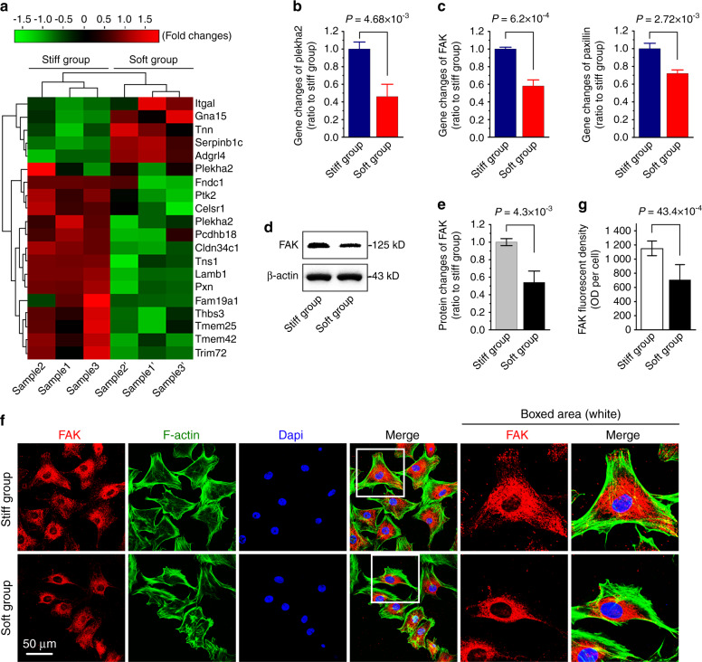
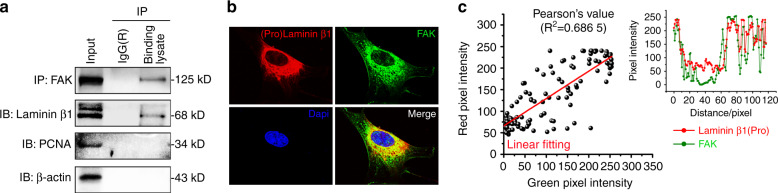
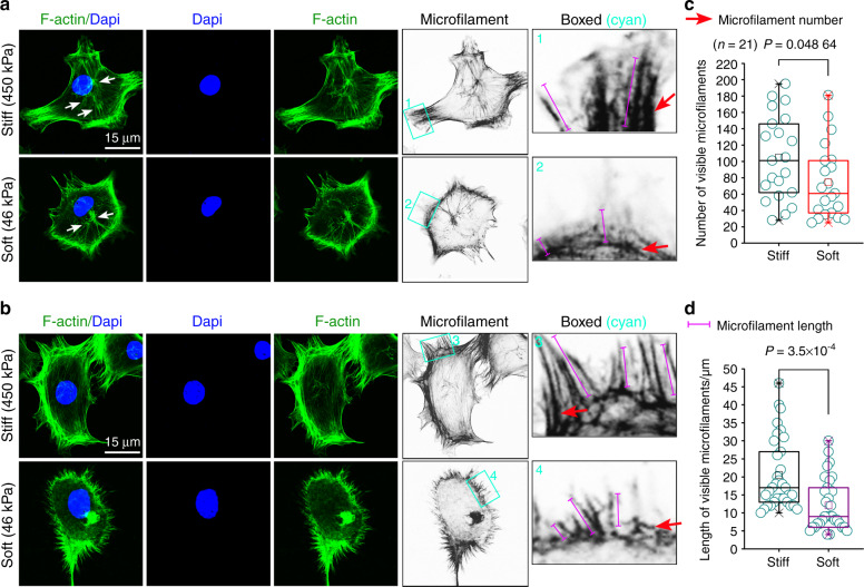
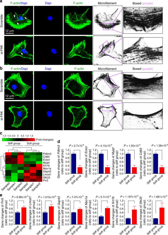
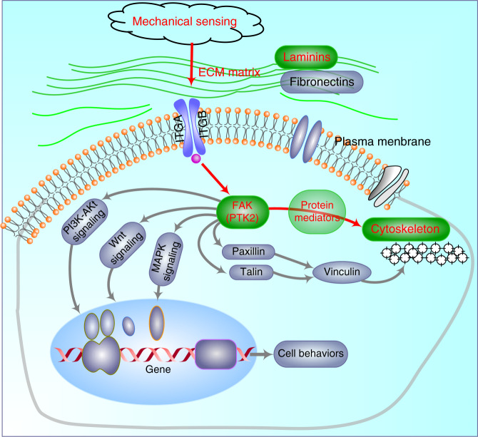
Microenvironmental biophysical factors play a fundamental role in controlling cell behaviors including cell morphology, proliferation, adhesion and differentiation, and even determining the cell fate. Cells are able to actively sense the surrounding mechanical microenvironment and change their cellular morphology to adapt to it. Although cell morphological changes have been considered to be the first and most important step in the interaction between cells and their mechanical microenvironment, their regulatory network is not completely clear. In the current study, we generated silicon-based elastomer polydimethylsiloxane (PDMS) substrates with stiff (15:1, PDMS elastomer vs. curing agent) and soft (45:1) stiffnesses, which showed the Young's moduli of ~450 kPa and 46 kPa, respectively, and elucidated a new path in cytoskeleton re-organization in chondrocytes in response to changed substrate stiffnesses by characterizing the axis shift from the secreted extracellular protein laminin β1, focal adhesion complex protein FAK to microfilament bundling. We first showed the cellular cytoskeleton changes in chondrocytes by characterizing the cell spreading area and cellular synapses. We then found the changes of secreted extracellular linkage protein, laminin β1, and focal adhesion complex protein, FAK, in chondrocytes in response to different substrate stiffnesses. These two proteins were shown to be directly interacted by Co-IP and colocalization. We next showed that impact of FAK on the cytoskeleton organization by showing the changes of microfilament bundles and found the potential intermediate regulators. Taking together, this modulation axis of laminin β1-FAK-microfilament could enlarge our understanding about the interdependence among mechanosensing, mechanotransduction, and cytoskeleton re-organization.


Conflict of interest statement

The authors declare no competing interests.

Figures

Fig. 1
Fig. 1
Cell morphology changes of chondrocytes in response to PDMS substrates with different stiffnesses. a Representative SEM images showing the cell morphology changes of chondrocytes seeded onto PDMS substrates with stiff (450 kPa) and soft (46 kPa) stiffnesses. Yellow arrows indicates cellular synapses. b Box and whisker plot showing the changes of cell spreading areas of chondrocytes seeded onto PDMS substrates with stiff (450 kPa) and soft (46 kPa) stiffnesses. Twenty-eight cells per group from eight independent experiments were used to calculate the cell spreading areas. c Box and whisker plot showing the changes of cellular synapses of chondrocytes seeded onto PDMS substrates with stiff (450 kPa) and soft (46 kPa) stiffnesses. Twenty-eight cells per group from eight independent experiments were used to calculate the cellular synapses. d Box and whisker plot showing the changes of cellular synapse lengths of chondrocytes seeded onto PDMS substrates with stiff (450 kPa) and soft (46 kPa) stiffnesses. Two hundred and eighty cellular synapses of twenty-eight cells from the stiff group and one hundred and twenty-five cellular synapses of twenty-eight cells from the stiff group were applied to calculate the cellular synapse lengths. The SEM experiments are based on eight independent experiments (n = 8). The data in (b), (c), & (d) are shown in the box (from 25%, 50% to 75%) and whisker (minimum to maximum values) plots. All significant data presented in (b), (c) & (d) are based on two-tailed Student’s t-tests.
Fig. 2
Fig. 2
Expressions of laminin β1 in chondrocytes in response to PDMS substrates with different stiffnesses. a RNA sequencing indicating the basal gene expression levels of laminin beta family in chondrocytes. Their expressions were all ratio to the inner β-actin gene. b qPCR showing mRNA changes of laminin β1 in chondrocytes in response to stiff and soft substrates. β-actin gene was used as the inner control. c Western blotting indicating the protein changes of laminin β1 in chondrocytes in response to stiff and soft substrates. d OD quantification confirming the protein changes of laminin β1 in chondrocytes in response to stiff and soft substrates. e Immunofluorescence by CLSM showing the expressions of laminin β1 in chondrocytes in response to stiff and soft substrates. *indicates the expressions of laminin β1 at the site of connections between the two cells. Arrows further indicate the details of laminin β1 at the site of connections between the two cells.The experiments in (a), (b), (c), & (e) are all based on three independent experiments (n = 3). The data in F are shown in the box (from 25%, 50% to 75%) and whisker (minimum to maximum values) plots. All significant data presented in (a), (b), (d), and (f) are based on two-tailed Student’s t-tests.
Fig. 3
Fig. 3
Expression profile of extracellular proteins/focal adhesion proteins in chondrocytes in response to PDMS substrates with different stiffnesses. a Pheatmap by RNA sequencing indicating the mRNA changes of extracellular proteins/focal adhesion proteins in chondrocytes in response to stiff and soft substrates. Cell layste samples were collected at 72 h after being seeded onto stiff and soft substrates. Data were presented as FPKM (Fragments Per Kilobase of transcript per Million fragments mapped) by online R-package from RNA sequencing data. Three independent pairs of samples, i.e., Sample 1 and 1’, Sample 2 and 2’, and Sample 3 and 3’, were from the same mother cells, respectively. b, c qPCR confirming the mRNA changes of plekha2, FAK and pxn in chondrocytes in response to stiff and soft substrates. β-actin gene was used as the inner control. d Western blotting showing the FAK protein in chondrocytes in response to stiff and soft substrates. e OD quantification confirming the protein changes of FAK in chondrocytes in response to stiff and soft substrates. f Immunofluorescence by CLSM showing the expressions of FAK in chondrocytes in response to stiff and soft substrates. g Fluorescent OD quantification confirming the protein changes of FAK in chondrocytes in response to stiff and soft substrates. The experiments in (b), (c), & (f) are all based on three independent experiments (n = 3). All significant data presented in (b), (c), (e), & (g) are based on two-tailed Student’s t-tests.
Fig. 4
Fig. 4
Interaction between the secreted extracellular protein laminin β1 and focal adhesion protein FAK. a Co-IP showing the binding between laminin β1 and FAK in chondrocytes. FAK was chosen to be the bait protein and laminin β1 was the prey protein. Nuclear protein PCNA (Proliferating cell nuclear antigen) was chosen to be a negative prey protein. β-actin was not only an internal control, but also a constituent unit of cytoskeleton. b Immunofluorescence by CLSM showing the co-expressions of laminin β1 (pro-protein) and FAK in chondrocytes. Laminin β1 was shown to be red fluorescent light and FAK was to be green fluorescent light. The objective lens was selected to be 100 × (Oil immersion lens). c Co-location analysis indicating the co-expressed relation between laminin β1 (pro-protein) and FAK in chondrocytes. The expression of co-distribution between laminin β1 (pro-protein) and FAK was achieved by linear fitting (Pearson’ R value). All experiments are based on three independent experiments (n = 3). The presentation of images in (a) & (b) are chosen as the representative images.
Fig. 5
Fig. 5
Detailed changes of cytoskeleton in chondrocytes in response to PDMS substrates with different stiffnesses. a, b Immunofluorescence by CLSM showing the expressions of F-actin in chondrocytes in response to stiff and soft substrates. White arrows indicated formation of microfilaments (F-actin bundle) near the nucleus. Grayscale images were used to indicate the changes of microfilaments in chondrocytes in response to stiff and soft substrates. Cyan boxed areas indicated the differences in microfilament number and length. c Quantitative analysis indicating the changes of microfilament numbers in chondrocytes in response to stiff and soft substrates. d Quantitative analysis indicating the changes of microfilament lengths in chondrocytes in response to stiff and soft substrates. The experiments of F-actin immunofluorescent staining by CLSM are based on at least 20 independent experiments (n = 20). The data in (c) & (d) are shown in the box (from 25%, 50% to 75%) and whisker (minimum to maximum values) plots. All significant data presented in (c) & (d) are based on two-tailed Student’s t-tests.
Fig. 6
Fig. 6
The potential protein mediators participated in re-organization of cytoskeleton in chondrocytes in response to PDMS substrates with different stiffnesses. a, b Immunofluorescence by CLSM showing the formation and distribution of microfilaments (F-actin bundle) in chondrocytes after reduction of FAK by siRNA interference. White arrows indicated formation of microfilaments near the nucleus. Grayscale images were used to indicate the changes of microfilaments in chondrocytes in response to stiff and soft substrates. Purple boxed areas indicated the differences in microfilament bundle. c Pheatmap by RNA sequencing indicating the mRNA changes of protein mediators in chondrocytes in response to stiff and soft substrates. Cell layste samples were collected at 72 h after being seeded onto stiff and soft substrates. Data were presented as FPKM (Fragments Per Kilobase of transcript per Million fragments mapped) by online R-package from RNA sequencing data. Three independent pairs of samples, i.e., Sample 1 and 1’, Sample 2 and 2’, and Sample 3 and 3’, were from the same mother cells, respectively. d qPCR confirming the mRNA down-regulation of protein mediators in chondrocytes in response to soft substrates relative to the stiff group. e qPCR confirming the mRNA up-regulation of protein mediators in chondrocytes in response to soft substrates relative to the stiff group. All experiments by qPCR in (d) & (e) are based on three independent experiments (n = 3). All significant data presented in (d) & (e) are based on two-tailed Student’s t-tests.
Fig. 7
Fig. 7
The schematic diagram showing the mediation axis from mechanical sensing, extracellular laminin β1 forming, focal adhesion protein FAK changing to cytoskeleton re-organization. Mechanical signals enter the chondrocytes to change the focal adhesion proteins, and then initiate the activation of signaling network, and finally regulate a variety of biological behaviors of the chondrocytes. The gray networks in the biomechanical regulation are involved but not presented in the current study; The red axis indicates the mediation process in the current study.

Similar articles

Cited by

References

    1. Vining KH, Mooney DJ. Mechanical forces direct stem cell behaviour in development and regeneration. Nat. Rev. Mol. Cell Biol. 2017;18:728–742. - PMC - PubMed
    1. Engler AJ, Sen S, Sweeney HL, Discher DE. Matrix elasticity directs stem cell lineage specification. Cell. 2006;126:677–689. - PubMed
    1. Papalazarou V, Salmeron-Sanchez M, Machesky LM. Tissue engineering the cancer microenvironment-challenges and opportunities. Biophys. Rev. 2018;10:1695–1711. - PMC - PubMed
    1. Adebowale K, et al. Enhanced substrate stress relaxation promotes filopodia-mediated cell migration. Nat. Mater. 2021;20:1290–1299. - PMC - PubMed
    1. Bonnans C, Chou J, Werb Z. Remodelling the extracellular matrix in development and disease. Nat. Rev. Mol. Cell Biol. 2014;15:786–801. - PMC - PubMed

Publication types