. 2022 Mar 11;13(1):1278.
doi: 10.1038/s41467-022-28811-w.

The structural basis for regulation of the glutathione transporter Ycf1 by regulatory domain phosphorylation

Affiliations

The structural basis for regulation of the glutathione transporter Ycf1 by regulatory domain phosphorylation

Nitesh Kumar Khandelwal et al. Nat Commun. .

Abstract

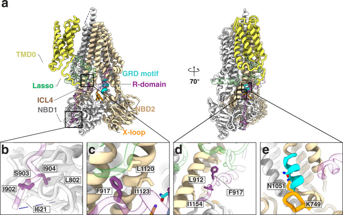
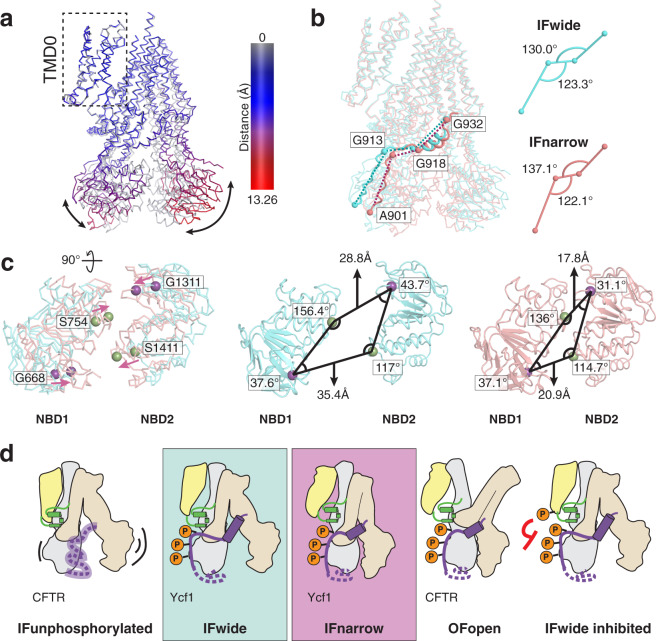
Yeast Cadmium Factor 1 (Ycf1) sequesters heavy metals and glutathione into the vacuole to counter cell stress. Ycf1 belongs to the ATP binding cassette C-subfamily (ABCC) of transporters, many of which are regulated by phosphorylation on intrinsically-disordered domains. The regulatory mechanism of phosphorylation is still poorly understood. Here, we report two cryo-EM structures of Ycf1 at 3.4 Å and 4.0 Å resolution in inward-facing open conformations that capture previously unobserved ordered states of the intrinsically disordered regulatory domain (R-domain). R-domain phosphorylation is clearly evident and induces a topology promoting electrostatic and hydrophobic interactions with Nucleotide Binding Domain 1 (NBD1) and the Lasso motif. These interactions stay constant between the structures and are related by rigid body movements of the NBD1/R-domain complex. Biochemical data further show R-domain phosphorylation reorganizes the Ycf1 architecture and is required for maximal ATPase activity. Together, we provide insights into how R-domains control ABCC transporter activity.


Conflict of interest statement

The authors declare no competing interests.

Figures

Fig. 1
Fig. 1. The regulatory architecture of Ycf1 determined by cryo-EM.
a Schematic of Ycf1 structural topology highlighting the arrangement of transmembrane helices belonging to transmembrane domain 0 (TMD0, yellow), transmembrane domain 1 (TMD1, gray), and transmembrane domain 2 (TMD2, wheat). Conserved cytoplasmic elements shown include the Lasso motif (green), the R-domain (purple), nucleotide binding domains 1 and 2 (NBD1 (gray); NBD2 (wheat)) and intracellular loops 1–4 (ICL1, ICL2 (dark gray); ICL3, ICL4 (brown)). Orange spheres corresponding to conserved putative phosphorylation sites in Ycf1 are labeled with their residue numbering on the corresponding domains. Cryo-EM model (left) and map (right) of the b IFwide (cyan) and c IFnarrow (pink) conformations of E1435Q Ycf1 colored using the same scheme as shown in (a). d ATPase activity in the presence of increasing concentrations of ATP for wild-type (WT) and the catalytically dead (E1435Q) variant of Ycf1. e Volume representation of the internal cavities of IFwide (left, cyan) and IFnarrow (right, pink). f ATPase activity in WT and E1435Q Ycf1 in the presence of increasing concentrations of oxidized glutathione (GSSG) and 1 mM ATP. Data are reported as stimulated rates in which the basal ATPase activity (without GSSG) was subtracted. Data shown correspond to a half-maximal effective concentration (EC50) of 8.7 ± 2.6 µM for GSSG-induced stimulation of ATPase activity in WT Ycf1. Results in (d) and (f) are the mean ± the standard deviation (SD) for n = 3 (technical triplicates). Source data are provided as a Source Data file.
Fig. 2
Fig. 2. The R-domain engages Ycf1 through an extensive phosphorylation-dependent network.
a Putative phosphorylation sites on the Ycf1 R-domain (purple) mapped onto a cartoon representation of the Ycf1 IFnarrow cryo-EM structure. b Sequence alignment of ABCC family members highlighting the consensuses sequences of putative phosphorylation motifs. Orange dots represent predicted phosphorylation sites. c Surface representation of the cryo-EM model of IFnarrow Ycf1 highlighting the orientation of the R-domain along NBD1. d A detailed view of the R-domain region shown in (c) highlighting residues contributing to the binding interface between the R-domain (purple), the Lasso motif (green), and NBD1 (gray). Carbon atoms are colored consistent with domain coloring in (c), with oxygen (red), nitrogen (blue), and phosphate (orange) atoms colored accordingly. Dashes represent hydrogen bonds or electrostatic interactions between heavy atoms. e Electron potential density of phosphorylated residues (S908, T911, and S914) observed in IFwide (cyan) and IFnarrow (pink) upper panel. SDS-PAGE analysis of phosphorylation in purified samples of Ycf1 in the presence or absence of Lambda phosphatase (Lambda PP) treatment. The left gel showing phosphorylated Ycf1 (pYcf1) was stained with Pro-Q phosphoprotein gel stain (Thermo Fisher), whereas the right gel was visualized with Coomassie gel stain to show total Ycf1 (Ycf1), Experiment is performed in biological replicate and representative gel is shown. f Representative SDS-PAGE analysis of proteolysis resistance in Lambda PP untreated (Ycf1) or treated (dephos-Ycf1) Ycf1 incubated with increasing concentrations of trypsin (0–20 µg/mL). The experiment was performed in biological replicate g ATPase activity of phosphorylated (Ycf1), dephosphorylated (dephos-Ycf1) and phosphorylated residue mutants (S908A, T911A, and S914A). Data shown are the mean ± SD for n = 4 (technical quadruplicates). Source data are provided as a Source Data file. h ATPase activity of the R716 mutant variant R716A and the R206 mutant variant R206E along with WT Ycf1. Data shown are the mean ± SD for n = 3 (technical triplicates). Source data are provided as a Source Data file.
Fig. 3
Fig. 3. R-domain interaction network in Ycf1.
a Overall structure of Ycf1 in IFnarrow. bd. Hydrophobic pockets along the R-domain/NBD interface. e Interactions between residues of the X-loop and GRD motif in IFnarrow. The R-domain is colored purple, TMD1-NBD1 is colored gray, TMD2 and NBD2 are colored wheat, Lasso motif is colored green, the X-loop is colored orange, and the GRD motif is colored cyan in all figures.
Fig. 4
Fig. 4. Proposed model for catalytic control in Ycf1 relies on transitions of the rigid R-domain architecture from IFwide to IFnarrow.
a Overlay of IFnarrow (gray) and IFwide states of Ycf1, in which IFwide is colored by RMSD calculated on a per residue basis from structural alignment between TMD0 of each state. b Comparison of the R-domain geometry and differences observed for the intradomain angles between flexible residues forming hinge regions in the R-domains in each state. c Superposed NBDs (left panel) from IFwide (cyan ribbon) and IFnarrow (pink ribbon) highlighting the rearrangement in the relative positions of conserved residues of the signature motif (S754 and S1411, green sphere) and the walker A motif (G668 and G1311, purple spheres) in each state. The specific interdomain angles formed by these sites in their respective states are shown in the middle (IFwide) and right (IFnarrow) panels. d Proposed model for organization and regulation of the R-domain through transport in light of the IFwide and IFnarrow Ycf1 structures and in the context of previously published CFTR structures (IFunphosphorylated (PDB ID: 5UAK); OFopen (PDB ID: 6MSM). Domains are colored yellow for TMD0, gray for TMD1 and NBD1, wheat for TMD2 and NBD2, green for the Lasso motif, and purple for the R-domain.

Similar articles

Cited by

References

    1. Gueldry O, et al. Ycf1p-dependent Hg(II) detoxification in Saccharomyces cerevisiae. Eur. J. Biochem. 2003;270:2486–2496. - PubMed
    1. Li ZS, et al. A new pathway for vacuolar cadmium sequestration in Saccharomyces cerevisiae: YCF1-catalyzed transport of bis(glutathionato)cadmium. Proc. Natl Acad. Sci. USA. 1997;94:42–47. - PMC - PubMed
    1. Szczypka MS, Wemmie JA, Moye-Rowley WS, Thiele DJ. A yeast metal resistance protein similar to human cystic fibrosis transmembrane conductance regulator (CFTR) and multidrug resistance-associated protein. J. Biol. Chem. 1994;269:22853–22857. - PubMed
    1. Paumi CM, Pickin KA, Jarrar R, Herren CK, Cowley ST. Ycf1p attenuates basal level oxidative stress response in Saccharomyces cerevisiae. FEBS Lett. 2012;586:847–853. - PMC - PubMed
    1. Morgan B, et al. Multiple glutathione disulfide removal pathways mediate cytosolic redox homeostasis. Nat. Chem. Biol. 2013;9:119–125. - PubMed

MeSH terms

Substances