

Accuracy of B-mode ultrasound and ARFI elastography in predicting malignancy of canine splenic lesions
- PMID: 35277580
- PMCID: PMC8917151
- DOI: 10.1038/s41598-022-08317-7
Accuracy of B-mode ultrasound and ARFI elastography in predicting malignancy of canine splenic lesions
Abstract
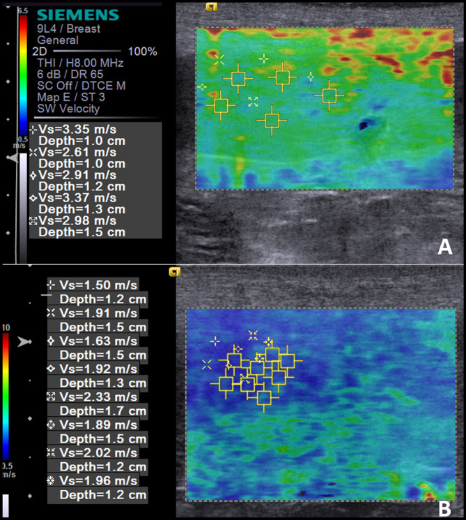
The objective was to evaluate the accuracy of B-mode ultrasonography and ARFI elastography in detecting malignancy in canine splenic lesions. Thirty-seven spleens with abnormalities (16 benign and 21 malignant) from dogs of different breeds and ages were evaluated. Echogenicity, echotexture, organ length and height were evaluated using B-mode. By ARFI elastography, tissue stiffness was evaluated qualitatively (elastogram) and quantitatively (measuring the shear wave velocity-SWV). Lesions were classified as diffuse, focal or multifocal (cranial, medial or caudal portion) and comparisons of the SWV between the injured and non-injured areas were performed. In the B-mode, no features were associated to malignancy (P > 0.05). In the elastogram, 35 spleens were non-deformable and 2 deformable, having no association with malignancy. The greater SWV was observed in malignant lesions (3.4 ± 0.6 m/s), followed by areas free from alterations (2.1 ± 0.3 m/s) and benign lesions (1.7 ± 0.5 m/s), with difference between groups (P < 0.0001). It was found that a SWV > 2.6 m/s indicates malignancy of canine splenic lesions (sensitivity of 95%, specificity of 100%, PPV of 100%, NPV of 94% and accuracy of 97%), concluding that ARFI elastography is a promising technique for differentiating malignancy in these lesions.
© 2022. The Author(s).
Conflict of interest statement
The authors declare no competing interests.
Figures
Similar articles
-
Ultrasonography methods for predicting malignancy in canine mammary tumors.PLoS One. 2017 May 22;12(5):e0178143. doi: 10.1371/journal.pone.0178143. eCollection 2017. PLoS One. 2017. PMID: 28542533 Free PMC article.
-
Malignancy prediction of cutaneous and subcutaneous neoplasms in canines using B-mode ultrasonography, Doppler, and ARFI elastography.BMC Vet Res. 2022 Jan 3;18(1):10. doi: 10.1186/s12917-021-03118-y. BMC Vet Res. 2022. PMID: 34980124 Free PMC article.
-
Diagnostic Value of ARFI (Acoustic Radiation Force Impulse) in Differentiating Benign From Malignant Breast Lesions.Acad Radiol. 2017 Jan;24(1):45-52. doi: 10.1016/j.acra.2016.09.001. Epub 2016 Oct 17. Acad Radiol. 2017. PMID: 27765598
-
Breast elastography: the technical process and its applications.Diagn Interv Imaging. 2013 May;94(5):503-13. doi: 10.1016/j.diii.2013.02.006. Epub 2013 Apr 22. Diagn Interv Imaging. 2013. PMID: 23619293 Review.
-
Quantitative Shear Wave Velocity Measurement on Acoustic Radiation Force Impulse Elastography for Differential Diagnosis between Benign and Malignant Thyroid Nodules: A Meta-analysis.Ultrasound Med Biol. 2015 Dec;41(12):3035-43. doi: 10.1016/j.ultrasmedbio.2015.08.003. Epub 2015 Sep 12. Ultrasound Med Biol. 2015. PMID: 26371402 Review.
Cited by
-
Shear Wave Elastography Evaluation of Testicular Stiffness in Dogs Affected by Testicular Pathology.Animals (Basel). 2025 Jan 26;15(3):353. doi: 10.3390/ani15030353. Animals (Basel). 2025. PMID: 39943123 Free PMC article.
-
In vivo endoscopic optical coherence elastography based on a miniature probe.Biomed Opt Express. 2024 Jun 12;15(7):4237-4252. doi: 10.1364/BOE.521154. eCollection 2024 Jul 1. Biomed Opt Express. 2024. PMID: 39022537 Free PMC article.
-
Diagnosis, Prognosis, and Treatment of Canine Hemangiosarcoma: A Review Based on a Consensus Organized by the Brazilian Association of Veterinary Oncology, ABROVET.Cancers (Basel). 2023 Mar 29;15(7):2025. doi: 10.3390/cancers15072025. Cancers (Basel). 2023. PMID: 37046686 Free PMC article. Review.
-
Two-Dimensional Shear-Wave Elastography of the Thyroid in Clinically Healthy Dogs in Different Age Groups.Animals (Basel). 2024 May 22;14(11):1528. doi: 10.3390/ani14111528. Animals (Basel). 2024. PMID: 38891575 Free PMC article.
-
B-Mode Ultrasonography and Acoustic Radiation Force Impulse Elastography in Evaluation of Urothelial Carcinoma in Dogs.Animals (Basel). 2025 Apr 26;15(9):1223. doi: 10.3390/ani15091223. Animals (Basel). 2025. PMID: 40362038 Free PMC article.
References
-
- Bandinelli MB, Pavarini SP, Oliveira EC, Gomes DC, Cruz CEF, Driemeier D. Estudo retrospectivo de lesões em baços de cães esplenectomizados: 179 casos. Pesq. Vet. Bras. 2011;31:697–701. doi: 10.1590/S0100-736X2011000800011. - DOI
-
- O'Byrne K, Hosgood G. Splenic mass diagnosis in dogs undergoing splenectomy according to breed size. Vet. Rec. 2019;184:620. - PubMed
Publication types
MeSH terms
LinkOut - more resources
Full Text Sources
Medical
