. 2022 Mar 11;5(1):228.
doi: 10.1038/s42003-022-03179-1.

Understanding the functional role of membrane confinements in TNF-mediated signaling by multiscale simulations

Affiliations

Understanding the functional role of membrane confinements in TNF-mediated signaling by multiscale simulations

Zhaoqian Su et al. Commun Biol. .

Abstract

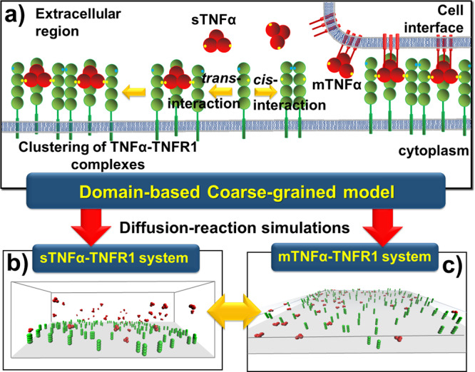
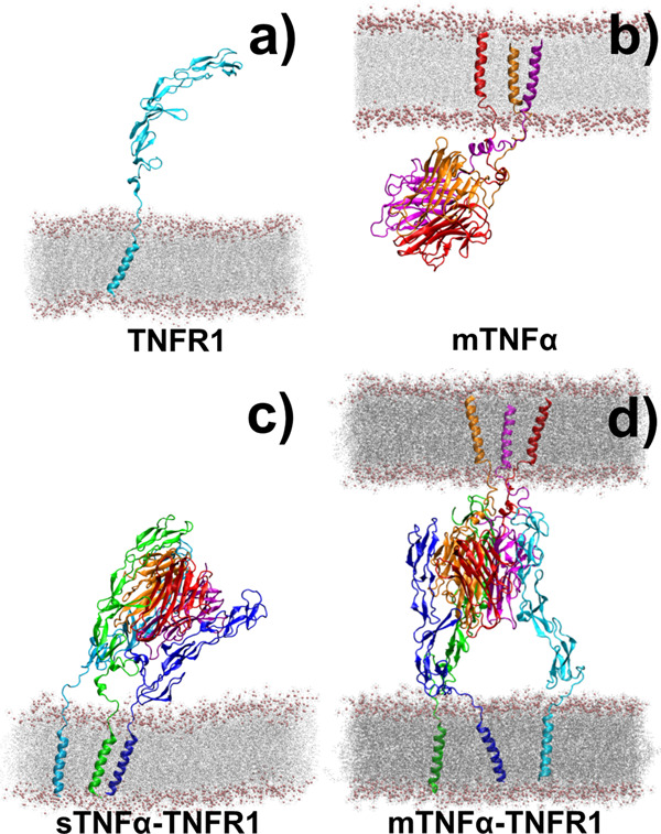
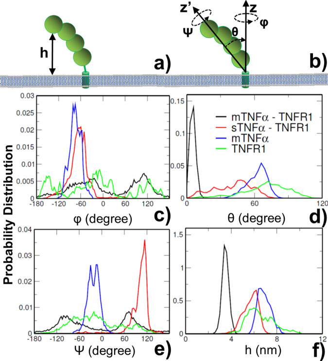
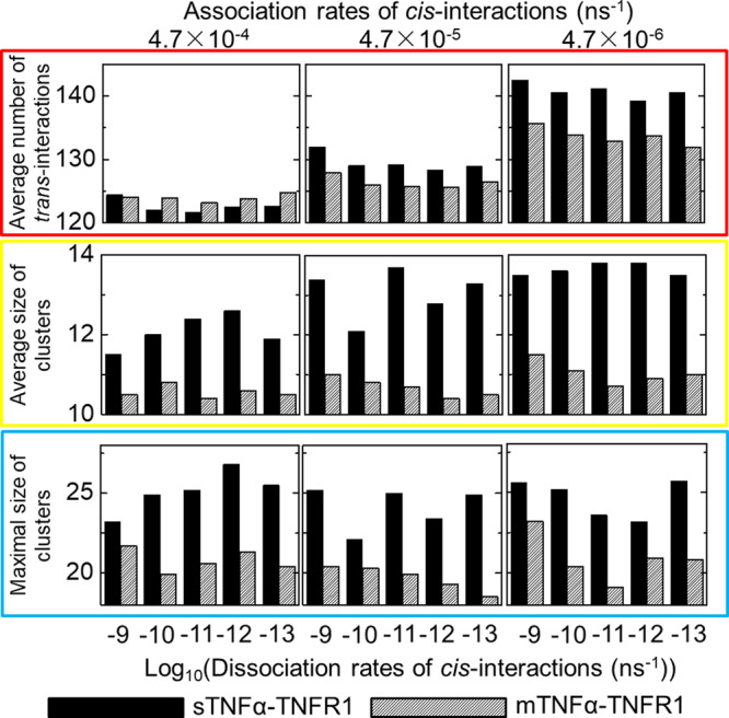
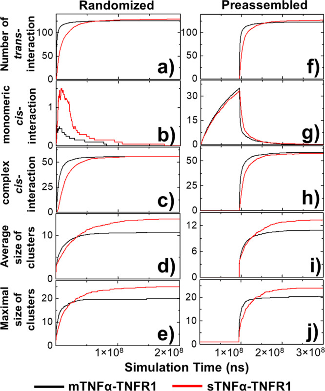
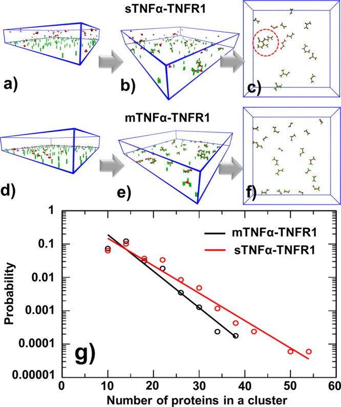
The interaction between TNFα and TNFR1 is essential in maintaining tissue development and immune responses. While TNFR1 is a cell surface receptor, TNFα exists in both soluble and membrane-bound forms. Interestingly, it was found that the activation of TNFR1-mediated signaling pathways is preferentially through the soluble form of TNFα, which can also induce the clustering of TNFR1 on plasma membrane of living cells. We developed a multiscale simulation framework to compare receptor clustering induced by soluble and membrane-bound ligands. Comparing with the freely diffusive soluble ligands, we hypothesize that the conformational dynamics of membrane-bound ligands are restricted, which affects the clustering of ligand-receptor complexes at cell-cell interfaces. Our simulation revealed that only small clusters can form if TNFα is bound on cell surface. In contrast, the clustering triggered by soluble TNFα is more dynamic, and the size of clusters is statistically larger. We therefore demonstrated the impact of membrane-bound ligand on dynamics of receptor clustering. Moreover, considering that larger TNFα-TNFR1 clusters is more likely to provide spatial platform for downstream signaling pathway, our studies offer new mechanistic insights about why the activation of TNFR1-mediated signaling pathways is not preferred by membrane-bound form of TNFα.


Conflict of interest statement

The authors declare no competing interests.

Figures

Fig. 1
Fig. 1. The multiscale simulation framework.
TNFα ligands (red) are initially presented on cell surfaces (mTNFα). They also occur as soluble variants (sTNFα) after their “stalk” regions are cleaved by metalloproteases (a). The trimeric ligands can simultaneously form “trans-interactions” with three receptors TNFR1 (green). Additionally, two TNFR1 receptors can also form a “cis-interaction”, through their PLAD regions (green dots) which do not spatially interfere with the trans-binding sites (yellow dots). It was found that the activation of TNFR1-mediated signaling pathways is more preferred by sTNFα than mTNFα, while both forms of ligands can further induce the aggregation of TNFR1 into nanoscale clusters through the combination of trans- and cis-interaction. Using a domain-based coarse-grained model and diffusion-reaction simulation algorithm, we compare the system in which clustering of TNFR1 is induced by soluble TNFα (b) to the system in which clustering is induced by membrane-bound TNFα (c).
Fig. 2
Fig. 2. The all-atom structural models of four specific cellular systems.
The conformational dynamics of ligand and receptor in these systems was analyzed by molecular dynamics simulations. The first system is the monomeric receptor TNFR1 on plasma membrane (a). The second system is the membrane-anchored TNFα ligand trimer (b). The third system is the complex formed between soluble ligand trimer and three membrane-bound TNFR1 receptors (sTNFα-TNFR1) (c). Finally, the last system is the complex formed between three membrane-bound TNFR1 receptors and a trimeric ligand TNFα which transmembrane regions are attached to an opposite plasma membrane (mTNFα-TNFR1) (d).
Fig. 3
Fig. 3. The distributions of conformational parameters derived from molecular dynamic simulations.
These parameters include: the translational fluctuations of proteins along membrane normal, h, as shown in a, and three Euler angles which characterize the rotational phase space, as illustrated in b. The distributions of the angle around the long principal axis z′ of the protein ψ are shown in c as indexed by curves with different colors. the distributions of the tilting angle between this principal axis and the membrane normal θ are shown in d; and the distribution of the angle around the membrane normal z φ are shown in e. Similarly, detailed distributions of translational fluctuations are shown in f for proteins in four modeled systems.
Fig. 4
Fig. 4. The results from domain-based diffusion-reaction simulations under different values of cis-interactions.
Because binding constants of cis-interactions between TNFR1 receptors have not been experimentally characterized, different combinations of association and dissociation rates were tested in the domain-based simulations. For each combination, 20 simulation trajectories were carried out. We calculated the average number of ligand-receptor trans-interactions (red frame), as well as the average (yellow frame) and maximal (blue frame) size of clusters obtained from each combination after all simulations were terminated. The left, middle and right columns indicate the specific values of association rate, while dissociation rates used in the simulations are indexed at the bottom of each plot. The data derived from sTNFα-TNFR1 and mTNFα-TNFR1 systems are represented by black and striped bars, respectively.
Fig. 5
Fig. 5. The comparison of kinetic profiles between simulations of soluble ligand and membrane-bound ligand.
We compare the kinetic profiles averaged from sTNFα-TNFR1 system (red) with mTNFα-TNFR1 (black) system as a function of simulation time. These profiles include the average numbers of trans-interactions (a) among different trajectories; the average numbers of monomeric (b) and ligand-bound cis-interactions (c) among different trajectories; and the average (d) and maximal (e) size of clusters. Moreover, we tested an alternative starting model in which the ligand-receptor interactions were turned off at the beginning so that monomeric TNFR1 receptors can preassemble. The kinetic profiles from this alternative starting model were also plotted as a function of simulation time, including the average numbers of trans-interactions (f) among different trajectories; the average numbers of monomeric (g) and ligand-bound cis-interactions (h) among different trajectories; and the average (i) and maximal (j) size of clusters.
Fig. 6
Fig. 6. The representative snapshots and cluster size distributions generated by domain-based diffusion-reaction simulations.
Some representative snapshots were selected from the simulations to visualize the spatial process of clustering. Specifically, the initial (a), middle (b) and final (c) configurations along a trajectory in sTNFα-TNFR1 system are compared with the initial (d), middle (e) and final (f) configurations along a trajectory in mTNFα-TNFR1 system. We found that large clusters can be organized into hexagonal lattice, as highlighted by the red dashed circle. Finally, we compared the cluster size distributions (g) in sTNFα-TNFR1 system (red) to the distribution in mTNFα-TNFR1 system (black). Given the logarithmic scale of the y-axis, the distributions of cluster size in both systems can be fitted by a single exponential function, whereas the clusters formed by sTNFα-TNFR1 complexes have the feasibility to grow into larger sizes.

Similar articles

Cited by

References

    1. Dostert C, Grusdat M, Letellier E, Brenner D. The TNF family of ligands and receptors: communication modules in the immune system and beyond. Physiol. Rev. 2019;99:115–160. - PubMed
    1. Wallach, D. The tumor necrosis factor family: family conventions and private idiosyncrasies. Cold Spring Harb. Perspect. Biol.10, a028431 (2018). - PMC - PubMed
    1. Aggarwal BB, Gupta SC, Kim JH. Historical perspectives on tumor necrosis factor and its superfamily: 25 years later, a golden journey. Blood. 2012;119:651–665. - PMC - PubMed
    1. Locksley RM, Killeen N, Lenardo MJ. The TNF and TNF receptor superfamilies: integrating mammalian biology. Cell. 2001;104:487–501. - PubMed
    1. MacEwan DJ. TNF ligands and receptors-a matter of life and death. Br. J. Pharm. 2002;135:855–875. - PMC - PubMed

Publication types