. 2022 Dec;25(4):895-904.
doi: 10.1007/s40477-022-00672-5. Epub 2022 Mar 12.

Treatment of canine and feline sarcoma using MR-guided focused ultrasound system

Affiliations

Treatment of canine and feline sarcoma using MR-guided focused ultrasound system

Anastasia Antoniou et al. J Ultrasound. 2022 Dec.

Abstract

Purpose: In recent years, veterinary medicine has enhanced its applications beyond traditional approaches, progressively incorporating the Focused Ultrasound (FUS) technology. This study investigated the ability of FUS to precisely ablate naturally occurring canine and feline soft tissue sarcomas (STS).

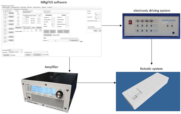
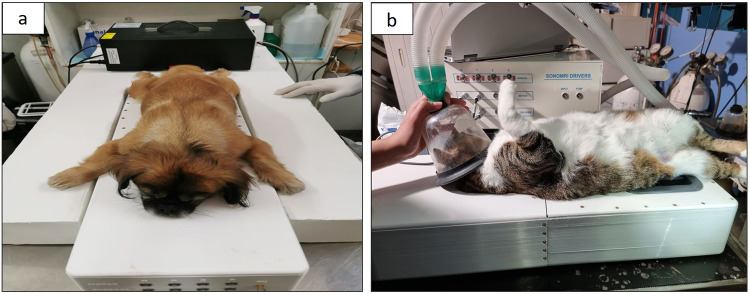
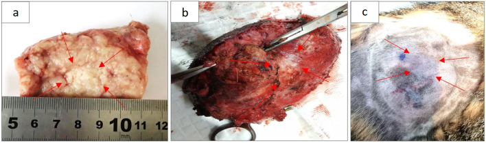
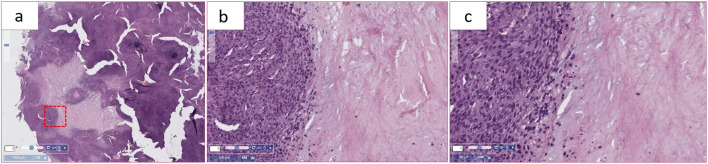
Methods: Six dogs and four cats with superficial tumours were enrolled in the study. The tumours were treated with a Magnetic Resonance guided FUS (MRgFUS) robotic system featuring a single element spherically focused transducer of 2.6 MHz. The tumours were then removed by surgery and sent for hematoxylin and eosin (H&E) staining.

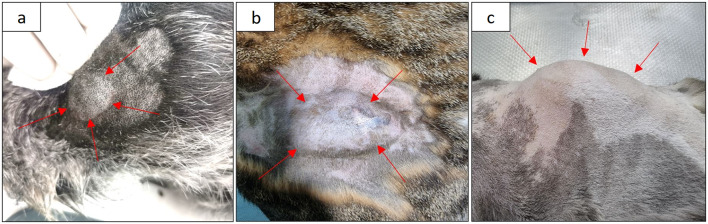
Results: The MRgFUS system was capable of inflicting well-defined overlapping lesions in the tumours. The anatomical sites of the treated tumours were the neck, leg, face, back and belly. Coagulative necrosis was evidenced by histopathology assessment in 80% of cases.

Conclusion: Therefore, this technology can be a therapeutic solution for veterinary cancer and a model for advancing the knowledge on human STS.

Keywords: Cats; Dogs; MRgFUS; Robotic device; Sarcomas; Superficial cancer.


Conflict of interest statement

All authors declare no conflict of interest.

Figures

Fig. 1
Fig. 1
Schematic diagram of the experimental set up
Fig. 2
Fig. 2
Placement of a a dog in prone position and a b cat in supine position on the robotic device at the veterinary premises
Fig. 3
Fig. 3
a Skin tumour located close to the eye of a dog and b–c Sarcoma in the back of a cat
Fig. 4
Fig. 4
Placement of a cat leg sarcoma above the ultrasonic transducer
Fig. 5
Fig. 5
a Lesion inflicted on skin tumour of a dog after single exposure (66 W acoustical power and 60 s pulse duration), b Overlapping lesions inflicted on skin tumour of a dog after ablation in a 2 × 2 grid pattern (3 mm step, 10 s pulse duration, 30 s delay, 66 W acoustical power, and 1 cm focal depth), and c Overlapping lesions inflicted on skin tumour of a cat after ablation in a 3 × 3 grid pattern (3 mm step, 10 s pulse duration, 30 s delay, 66 W acoustical power, and 1 cm focal depth) using the 2.6 MHz transducer (with 40 mm diameter and 60 mm radius of curvature)
Fig. 6
Fig. 6
Histological slides of a cat sarcoma after ablation in 3 × 3 grid (3 mm step, 10 s pulse duration, 30 s delay, 49.5 W acoustical power, and 2 cm focal depth) using a 10X and b 20X magnification. Blue arrows indicate a marked area of coagulative necrosis and red arrows indicates blood coagulation at the lesion borderline
Fig. 7
Fig. 7
Histological slides of a dog skin tumour revealing necrotic area resulted from ablation in 3 × 3 grid (3 mm step, 10 s pulse duration, 30 s delay, 66 W acoustical power, 1 cm focal depth) using a no magnification, b 10X, and c 20X magnification

References

    1. Davis BW, Ostrander EA. Domestic dogs and cancer research: a breed-based genomics approach. ILAR J. 2014;55(1):59–68. doi: 10.1093/ilar/ilu017. - DOI - PMC - PubMed
    1. Theilen GH and Madewell BR (1979) Tumors of the skin and subcutaneous tissues. 1st Philad., G. H. Theilen and B. R. Madewell, Eds. Veterinary Cancer Medicine 123–191
    1. Dobson JM, Samuel S, Milstein H, Rogers K, Wood JLN. Canine neoplasia in the UK: estimates of incidence rates from a population of insured dogs. J Small Anim Pract. 2002;43(6):240–246. doi: 10.1111/j.1748-5827.2002.tb00066.x. - DOI - PubMed
    1. Bray JP. Soft tissue sarcoma in the dog—part 1: a current review. J Small Anim Pract. 2016;57(10):510–519. doi: 10.1111/jsap.12556. - DOI - PubMed
    1. Hohenhaus AE, et al. Canine cutaneous and subcutaneous soft tissue sarcoma: an evidence-based review of case management. J Am Anim Hosp Assoc. 2016;52(2):77–89. doi: 10.5326/JAAHA-MS-6305. - DOI - PubMed