. 2022 Apr 29;40(4):423-434.
doi: 10.1093/stmcls/sxac006.

Mechanically Induced Nuclear Shuttling of β-Catenin Requires Co-transfer of Actin

Affiliations

Mechanically Induced Nuclear Shuttling of β-Catenin Requires Co-transfer of Actin

Buer Sen et al. Stem Cells. .

Abstract

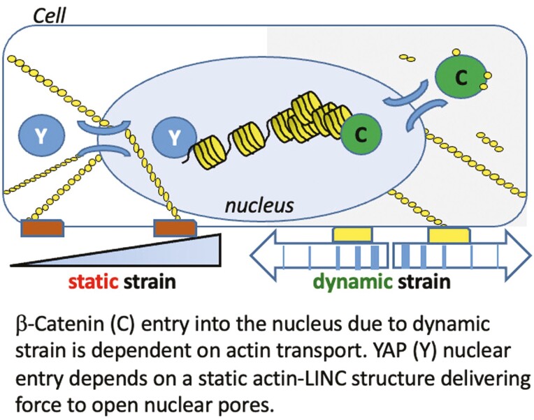
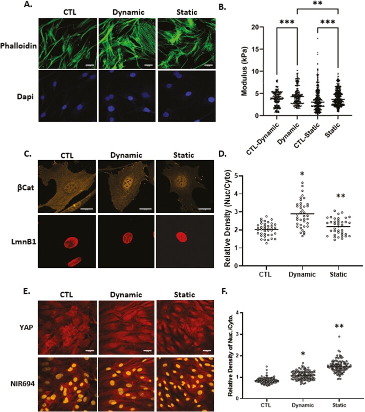
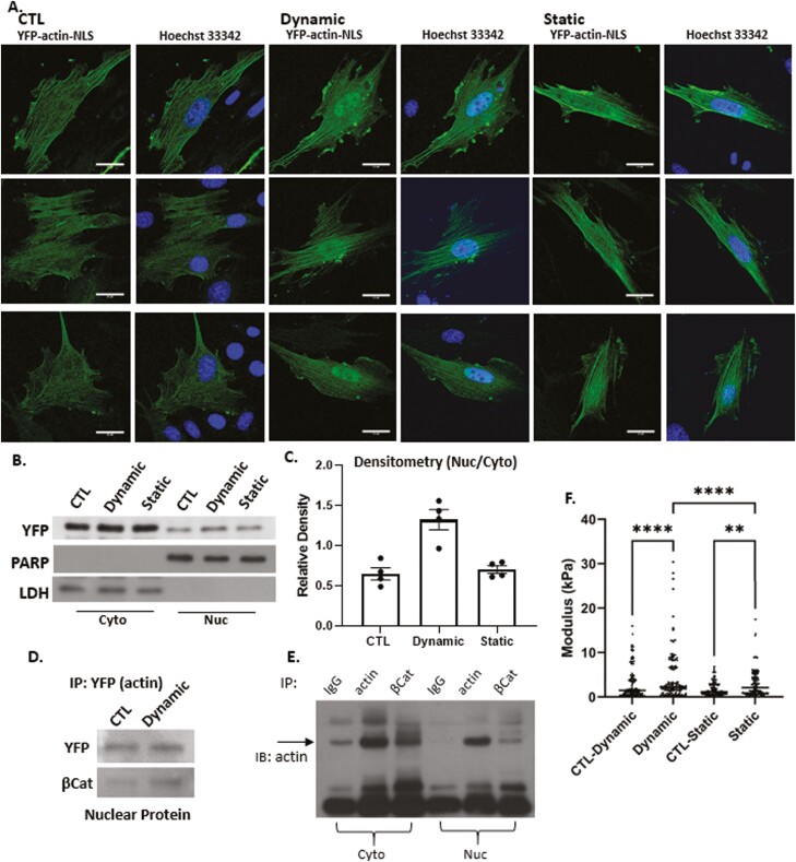
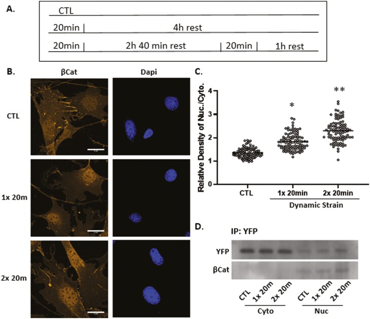
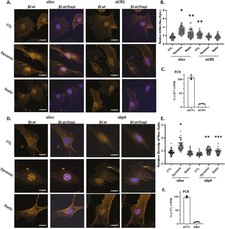
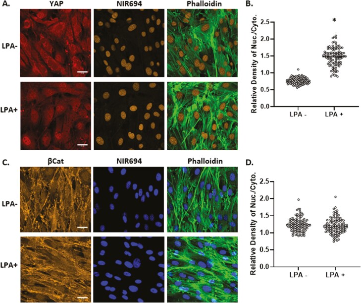
Mesenchymal stem cells (MSCs) respond to environmental forces with both cytoskeletal re-structuring and activation of protein chaperones of mechanical information, β-catenin, and yes-associated protein 1 (YAP1). To function, MSCs must differentiate between dynamic forces such as cyclic strains of extracellular matrix due to physical activity and static strains due to ECM stiffening. To delineate how MSCs recognize and respond differently to both force types, we compared effects of dynamic (200 cycles × 2%) and static (1 × 2% hold) strain on nuclear translocation of β-catenin and YAP1 at 3 hours after force application. Dynamic strain induced nuclear accumulation of β-catenin, and increased cytoskeletal actin structure and cell stiffness, but had no effect on nuclear YAP1 levels. Critically, both nuclear actin and nuclear stiffness increased along with dynamic strain-induced β-catenin transport. Augmentation of cytoskeletal structure using either static strain or lysophosphatidic acid did not increase nuclear content of β-catenin or actin, but induced robust nuclear increase in YAP1. As actin binds β-catenin, we considered whether β-catenin, which lacks a nuclear localization signal, was dependent on actin to gain entry to the nucleus. Knockdown of cofilin-1 (Cfl1) or importin-9 (Ipo9), which co-mediate nuclear transfer of G-actin, prevented dynamic strain-mediated nuclear transfer of both β-catenin and actin. In sum, dynamic strain induction of actin re-structuring promotes nuclear transport of G-actin, concurrently supporting nuclear access of β-catenin via mechanisms used for actin transport. Thus, dynamic and static strain activate alternative mechanoresponses reflected by differences in the cellular distributions of actin, β-catenin, and YAP1.

Keywords: YAP-signaling protein; actin cytoskeleton; mechanical stress; mesenchymal stem cell; nuclear transport.


Figures

None
The symmetry breaking of autophagic activity in CD133-positive human neuroblastoma cells. During cytokinesis, the recycling endosomal CD133 localizes to the pericentrosomal region asymmetrically to suppress autophagy. The asymmetric distribution of pericentrosomal CD133 endosomes then promotes the localization of β-catenin to the nucleus. β-catenin directly repressed p62/SQSTM1 expression. This suggests that pericentrosomal CD133 and β-catenin cooperatively suppress autophagy. In addition, nuclear β-catenin may inhibit pericentrosomal CD133 degradation to repress p62 gene expression.
Figure 1.
Figure 1.
Dynamic and static strains both increase cell stiffness yet have different effects on mechanoresponders. Mesenchymal stem cells were seeded in minimal essential medium at a density of 2000 or 10 000 cells/cm2 for β-catenin or YAP1 experiments respectively. Twenty-four hours later, application of 2% dynamic (200 cycles over 20 minutes) or static strain (2% and hold tension) was followed by staining with phalloidin (green), anti-β-catenin (yellow), or anti-YAP (red), scale bar = 25 μm (A, D, and E). Nuclear intensity of β-catenin and YAP1 was analyzed by ImageJ program and statistical significance indicated by letter a and b, both of them P < .0001 and ∗ ≠ ∗∗ (D and F). (b) Application of dynamic strain increased the AFM-measured cell modulus by 22% (P < .001, N = 350/grp) compared with non-strained control and while static strain resulted in a 13% increase compared with non-strained control (P < .0001, N = 800/grp). Cell modulus of MSCs treated with dynamic strain were, on average, 11% larger compared with static strain groups (P < .05).
Figure 2.
Figure 2.
Nuclear actin increases after dynamic strain. Nuclear YFP-NLS-βActin in MSC increases after dynamic, but not static strain (3 examples of each condition shown, scale bar = 25 μm (A). Western blot of cytoplasmic and nuclear lysates (B) was quantified by densitometry n = 4, P < .005 (C). IP nuclear protein using anti-GFP and IB YFP and β-catenin shows association between the 2 molecules (D). Cell cytoplasmic and nuclear lysates were IP for IgG, actin or βCat and immunoblotted for actin (E). AFM-measured modulus of isolated nuclei showed that dynamic strain increased the nuclear modulus by 2-fold (P < .001) compared to control and 1.3-fold compared with static strain groups (P < .001). Static strain increased nuclear modulus by 1.5-fold compared with control (P < .05) (F).
Figure 3.
Figure 3.
Nuclear β-catenin and actin rise further after a second dynamic strain bout. Timed application of strain for signal and double bouts (A). Mesenchymal stem cells were stained with anti-β-catenin (yellow) or DAPI (blue) showing increased β-catenin signal after the second strain bout, scale bar = 25 μm (B). β-catenin was quantified using ImageJ; statistical significance indicated by letter a and b, both = P < .0001 and ∗ ≠ ∗∗ (C). Nuclear and cytoplasmic fractions were pulled down with GFP Ab, and Western as shown for YFP and β-catenin, both of which increase after strain application (D).
Figure 4.
Figure 4.
β-Catenin nuclear entry is dependent on active actin transport. Cofilin-1 or importin-9 were knocked down in MSC prior to treatment with dynamic or static strain. Three hours later, cells were stained with anti-β-catenin (yellow) and DAPI (blue) (A and D), scale bar = 25 μm. Nuclear intensity of β-catenin was analyzed by ImageJ program and statistical significance indicated by letter a, b, or c. (i). ∗ ≠ ∗∗, P < .0001; (ii). ∗∗ ≠ ∗∗∗; P < .05 (B and E). Real-time PCR indicated knock-down of Cfl1 or Ipo9 (C and F).
Figure 5.
Figure 5.
YAP1, but not β-catenin, responds to generation of cytoplasmic F-actin. Mesenchymal stem cells at 10 000 cells/cm2 were treated with LPA (50 μM) for 24 hours and stained for YAP1 (red), nucleus (yellow), and F-actin (green) (A) or βCat (yellow), nucleus (blue), and F-actin (green), scale bar = 25 μm (A, C). Nuclear/cytoplasmic density of YAP1 or β-catenin IF was quantitated by ImageJ, ∗ = P < .0001 (B, D).

References

    1. Sen B, Xie Z, Case N, et al. Mechanical strain inhibits adipogenesis in mesenchymal stem cells by stimulating a durable beta-catenin signal. Endocrinology. 2008;149:6065-6075. 10.1210/en.2008-0687. - DOI - PMC - PubMed
    1. Dupont S, Morsut L, Aragona M, et al. Role of YAP/TAZ in mechanotransduction. Nature. 2011;474:179-183. 10.1038/nature10137. - DOI - PubMed
    1. Wang S, Englund E, Kjellman P, et al. CCM3 is a gatekeeper in focal adhesions regulating mechanotransduction and YAP/TAZ signalling. Nat Cell Biol. 2021;23:758-770. 10.1038/s41556-021-00702-0. - DOI - PubMed
    1. Sen B, Styner M, Xie Z, et al. Mechanical loading regulates NFATc1 and beta-catenin signaling through a GSK3beta control node. J Biol Chem. 2009;284:34607-34617. 10.1074/jbc.M109.039453. - DOI - PMC - PubMed
    1. Lara-Castillo N, Kim-Weroha NA, Kamel MA, et al. In vivo mechanical loading rapidly activates beta-catenin signaling in osteocytes through a prostaglandin mediated mechanism. Bone. 2015;76:58-66. 10.1016/j.bone.2015.03.019. - DOI - PMC - PubMed

Publication types

MeSH terms