Review
. 2022 Nov;12(11):1939-1957.
doi: 10.1002/2211-5463.13394. Epub 2022 Mar 22.

Neuronal SNARE complex assembly guided by Munc18-1 and Munc13-1

Affiliations
Review

Neuronal SNARE complex assembly guided by Munc18-1 and Munc13-1

Shen Wang et al. FEBS Open Bio. 2022 Nov.

Abstract

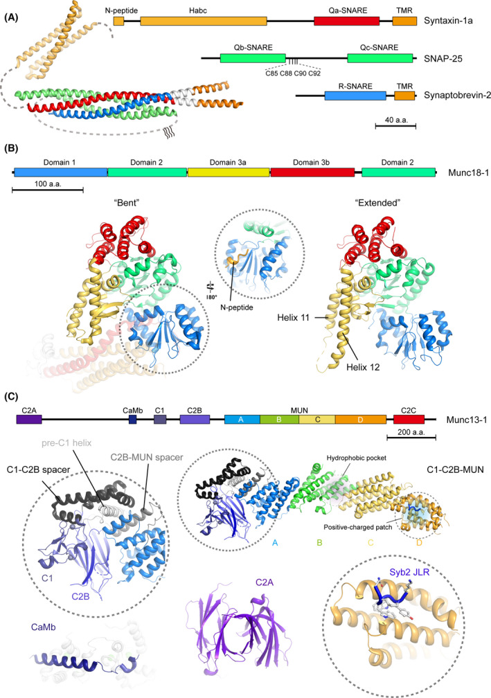
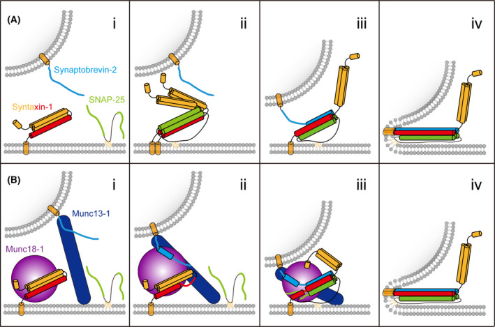
Neurotransmitter release by Ca2+ -triggered synaptic vesicle exocytosis is essential for information transmission in the nervous system. The soluble N-ethylmaleimide sensitive factor attachment protein receptors (SNAREs) syntaxin-1, SNAP-25, and synaptobrevin-2 form the SNARE complex to bring synaptic vesicles and the plasma membranes together and to catalyze membrane fusion. Munc18-1 and Munc13-1 regulate synaptic vesicle priming via orchestrating neuronal SNARE complex assembly. In this review, we summarize recent advances toward the functions and molecular mechanisms of Munc18-1 and Munc13-1 in guiding neuronal SNARE complex assembly, and discuss the functional similarities and differences between Munc18-1 and Munc13-1 in neurons and their homologs in other intracellular membrane trafficking systems.

Keywords: Munc13; Munc18; SNARE complex assembly; SNAREs; synaptic exocytosis; synaptic vesicle fusion.


Conflict of interest statement

The authors declare no conflict of interest.

Figures

Fig. 1
Fig. 1
Structure illustration of the neuronal SNAREs, Munc18‐1, and Munc13‐1. (A) Crystal structure and domain illustration of the neuronal SNARE complex. The structure models are fetched from the protein data bank (PDB) by entries of 3HD7 (helical extended neuronal SNARE complex) and 1EZ3 (Habc domain of syntaxin‐1). The SNARE motif of syntaxin‐1 (Qa‐SNARE, residues 191−253), SNAP‐25 (Qb‐ and Qc‐SNARE, residues 19−81 and 140−202), and synaptobrevin‐2 (R‐SNARE, residues 29−87) are colored in red, green, and blue, respectively. The N‐peptide (residues 1−10) and Habc (residues 26−146) of syntaxin‐1 are colored in bright orange. Transmembrane domains (TMRs) of syntaxin‐1 (residues 266−288) and synaptobrevin‐2 (residues 95−114) are colored in orange. The juxtamembrane linker regions (JLRs) of syntaxin‐1 and synaptobrevin‐2 are colored in light gray. The S‐palmitoyl cysteines of SNAP‐25 are indicated as C85, C88, C90, and C92, respectively. Missing densities in the structural model are supplied by dashed lines. (B) Structural illustration of Rattus norvegicus Munc18‐1 with two conformations. The subdomains of Munc18‐1 are colored in blue (domain 1, residues 4−134), green (domain 2, residues 135−245 and 490−592), yellow (domain 3a, residues 246−360), and red (domain 3b, residues 361−479), respectively. Left panel displays the ‘bent’ conformation of Munc18‐1 that binds to syntaxin‐1 (PDB entry: 3C98), where the helix 11 and 12 of domain 3a are folded back. Inset shows the binding between N‐peptide (bright orange) of syntaxin‐1 and domain 1 of Munc18‐1. Right panel displays the ‘extended’ conformation of Munc18‐1 (PDB entry: 3PUJ), in which helix 11 and 12 are outstretched. (C) Structural illustration of Rattus norvegicus Munc13‐1. The domains and subdomains of Munc13‐1 are colored in purple (C2A, residues 1−97), dark blue (calmodulin‐binding domain, CaMb, residues 459−492), navy blue (C1, residues 566−616), purple blue (C2B, residues 683−820), blue (MUN‐A, residues 859−1005), green (MUN‐B, residues 1006−1167), yellow (MUN‐C, residues 1168−1318), bright orange (MUN‐D, residues 1319−1531), and red (C2C, residues 1558−1685), respectively. The structural models of C1‐C2B‐MUN fragment (PDB entry: 5UE8 and 6A30), C2A (dimer form, PDB entry: 2CJT), and CaMb (binding with calmodulin, PDB entry: 2KDU) are fetched from PDB. Two functional regions of the MUN domain, that is, the hydrophobic pocket (binds to syntaxin‐1) and the negative‐charged patch (binds to synaptobrevin‐2), are highlighted by purple and blue shadows, respectively. Inset on the left shows the architecture of the intramolecule contacts of C1, C2B, and MUN domain. Inset on the right shows the interaction between the MUN‐D and synaptobrevin‐2 (Syb2) JLR.
Fig. 2
Fig. 2
Models of SNARE‐mediated membrane fusion and synaptic exocytosis. (A) The zippering model with merely the three neuronal SNAREs. (i) At rest state, syntaxin‐1 adopts self‐inhibitory conformation; (ii) syntaxin‐1 fluctuates between closed and open conformations and is prone to form a 2 : 1 complex with SNAP‐25; (iii) synaptobrevin‐2 displaces one copy of syntaxin‐1 of the 2 : 1 complex; the N‐termini of the SNARE motifs nucleate together to promote complex assembly; (iv) zippering of the SNARE motifs transfers sufficient energy into the membrane thus catalyzing membrane fusion. (B) Munc18‐1 and Munc13‐1 synergistically organize neuronal SNARE complex assembly and synaptic exocytosis. (i) Munc18‐1 captures syntaxin‐1 into closed conformation; Munc13‐1 bridges the presynaptic membrane and synaptic vesicle to facilitate synaptic vesicle docking; (ii) Munc13‐1 interacts with Munc18‐1−syntaxin‐1 complex to induce conformational changes of the syntaxin‐1 linker region and Munc18‐1 domain 3a, leading to uncaging of the N‐terminus of the syntaxin‐1 SNARE motif and extension of Munc18‐1 domain 3a; in the meantime, Munc18‐1 interacts with the C‐terminal half of synaptobrevin‐2 with the assistance of the binding between Munc13‐1 and synaptobrevin‐2 JLR. This intermediate underlies a potential conformational state, namely the prefusion priming complex; (iii) the N‐termini of the SNARE motifs start to nucleate to produce a half‐zippered SNARE complex, which is organized by Munc18‐1 and Munc13‐1; (iv) full zippering of the SNARE motifs in response to calcium signal is accompanied by the interplay of complexins/synaptotagmin‐1 with the half‐zippered SNARE complex (not shown). The color schemes of the neuronal SNAREs are the same as in Fig. 1. Munc18‐1 and Munc13‐1 are colored in purple and navy blue, respectively.

References

    1. Sudhof TC, Rizo J. Synaptic vesicle exocytosis. Cold Spring Harb Perspect Biol. 2011;3:a005637. 10.1101/cshperspect.a005637 - DOI - PMC - PubMed
    1. Sudhof TC. Neurotransmitter release: the last millisecond in the life of a synaptic vesicle. Neuron. 2013;80:675–90. 10.1016/j.neuron.2013.10.022 - DOI - PMC - PubMed
    1. Jahn R, Fasshauer D. Molecular machines governing exocytosis of synaptic vesicles. Nature. 2012;490:201–7. 10.1038/nature11320 - DOI - PMC - PubMed
    1. Emperador‐Melero J, Kaeser PS. Assembly of the presynaptic active zone. Curr Opin Neurobiol. 2020;63:95–103. 10.1016/j.conb.2020.03.008 - DOI - PMC - PubMed
    1. Rosenmund C, Rettig J, Brose N. Molecular mechanisms of active zone function. Curr Opin Neurobiol. 2003;13:509–19. 10.1016/j.conb.2003.09.011 - DOI - PubMed

Publication types

MeSH terms