

A Prospective Longitudinal Study to Investigate Corneal Hysteresis as a Risk Factor of Central Visual Field Progression in Glaucoma
- PMID: 35278360
- PMCID: PMC10249485
- DOI: 10.1016/j.ajo.2022.02.025
A Prospective Longitudinal Study to Investigate Corneal Hysteresis as a Risk Factor of Central Visual Field Progression in Glaucoma
Abstract
Purpose: To evaluate the role of corneal hysteresis (CH) as a risk factor of central visual field (VF) progression in a cohort of glaucoma suspect and glaucoma patients.
Design: Prospective cohort study.
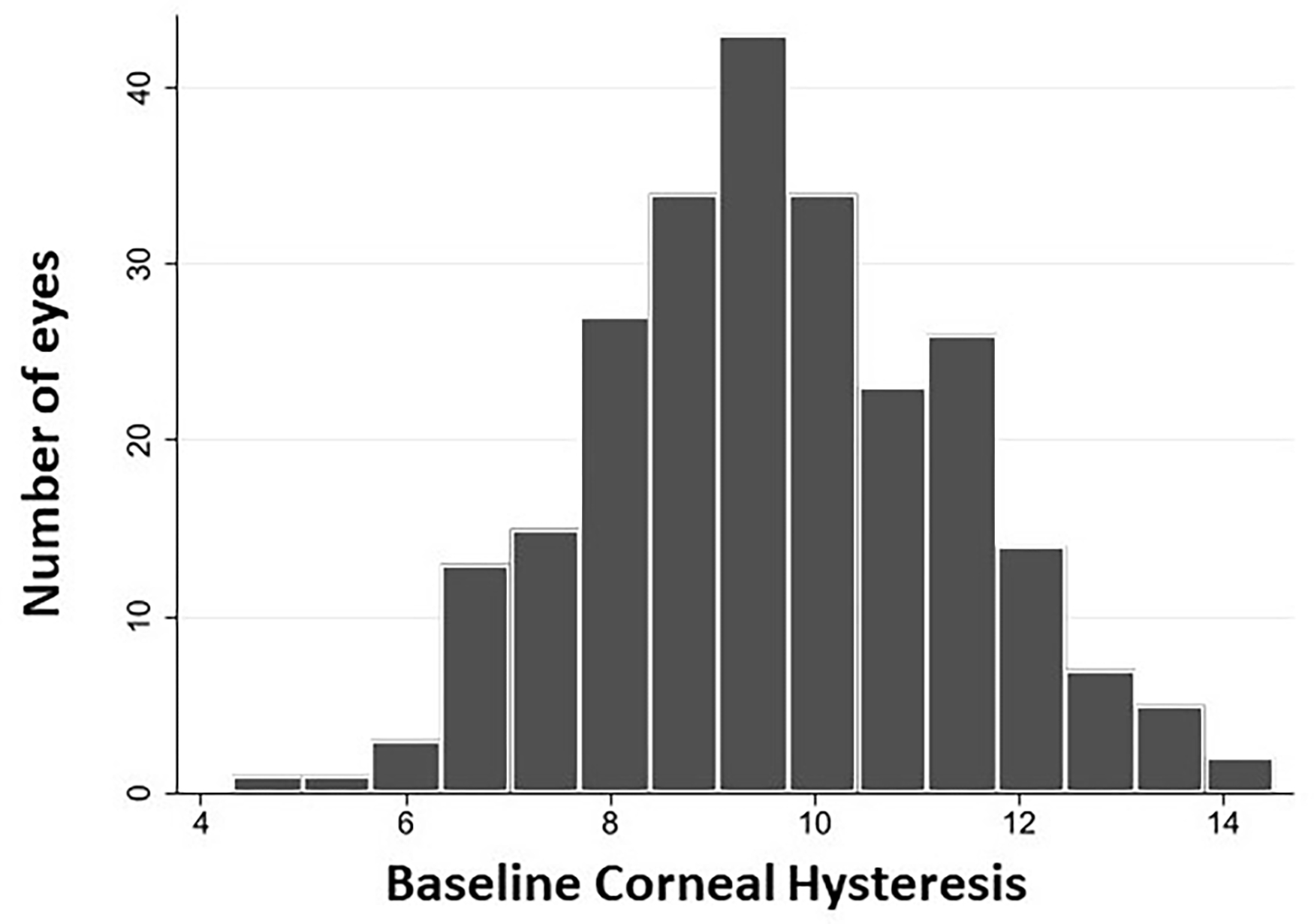
Methods: Two hundred forty-eight eyes of 143 subjects who were followed for an average of 4.8 years with a minimum of 5 visits with 10-2 and 24-2 VF tests were included. Univariable and multivariable linear mixed-effects models were used to identify characteristics associated with the rate of change over time in 10-2 and 24-2 mean deviation (MD). Mixed-effects logistic regression was used to evaluate characteristics associated with an increased likelihood of event-based 10-2 VF progression based on the clustered pointwise linear regression criterion.
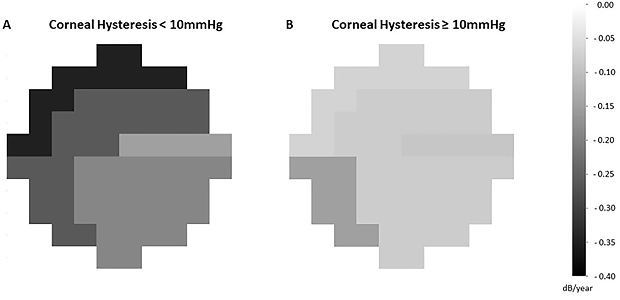
Results: CH was significantly associated with 10-2 and 24-2 VF progression in the univariable trend-based analysis. In multivariable trend-based analyses, lower CH was associated with a faster rate of decline in 10-2 MD (0.07 dB/y per 1 mm Hg, P < .001) but not with 24-2 MD (P = .490). In multivariable event-based analysis, lower CH was associated with an increased likelihood of 10-2 VF progression (odds ratio = 1.35 per 1 mm Hg lower, P = .025). Similar results were found in eyes with early glaucomatous damage at the baseline (baseline: 24-2 MD ≥ -6 dB).
Conclusions: Lower CH was associated with a statistically significant, but relatively small, increased risk of central VF progression on the 10-2 test grid. Given the substantial influence of central VF impairment on the quality of life, clinicians should consider using CH to assess the risk of progression in patients with primary open-angle glaucoma including those with early disease.
Copyright © 2022 Elsevier Inc. All rights reserved.
Figures
Similar articles
-
Central visual field in glaucoma: An updated review.Taiwan J Ophthalmol. 2024 Sep 13;14(3):360-370. doi: 10.4103/tjo.TJO-D-24-00042. eCollection 2024 Jul-Sep. Taiwan J Ophthalmol. 2024. PMID: 39430344 Free PMC article. Review.
-
Multilayer Macula Vessel Density and Visual Field Progression in Glaucoma.Am J Ophthalmol. 2022 May;237:193-203. doi: 10.1016/j.ajo.2021.11.018. Epub 2021 Nov 19. Am J Ophthalmol. 2022. PMID: 34801510 Free PMC article.
-
Baseline Age and Mean Deviation Affect the Rate of Glaucomatous Vision Loss.J Glaucoma. 2020 Jan;29(1):31-38. doi: 10.1097/IJG.0000000000001401. J Glaucoma. 2020. PMID: 31688371
-
Corneal hysteresis as a risk factor for glaucoma progression: a prospective longitudinal study.Ophthalmology. 2013 Aug;120(8):1533-40. doi: 10.1016/j.ophtha.2013.01.032. Epub 2013 May 1. Ophthalmology. 2013. PMID: 23642371 Free PMC article.
-
Biomechanical Glaucoma Factor and Corneal Hysteresis in Treated Primary Open-Angle Glaucoma and Their Associations With Visual Field Progression.Invest Ophthalmol Vis Sci. 2021 Jun 1;62(7):4. doi: 10.1167/iovs.62.7.4. Invest Ophthalmol Vis Sci. 2021. PMID: 34086046 Free PMC article.
Cited by
-
In vivo assessment of the ocular biomechanical properties in patients with idiopathic normal pressure hydrocephalus.Int Ophthalmol. 2024 Feb 2;44(1):1. doi: 10.1007/s10792-024-02922-3. Int Ophthalmol. 2024. PMID: 38315313 Free PMC article.
-
Central visual field in glaucoma: An updated review.Taiwan J Ophthalmol. 2024 Sep 13;14(3):360-370. doi: 10.4103/tjo.TJO-D-24-00042. eCollection 2024 Jul-Sep. Taiwan J Ophthalmol. 2024. PMID: 39430344 Free PMC article. Review.
-
Intereye Differences in the Clinical Assessment of Intraocular Pressure and Ocular Biomechanics.Optom Vis Sci. 2023 Oct 1;100(10):688-696. doi: 10.1097/OPX.0000000000002066. Epub 2023 Aug 29. Optom Vis Sci. 2023. PMID: 37639554 Free PMC article.
-
Intraocular pressure increases the rate of macular vessel density loss in glaucoma.Br J Ophthalmol. 2024 Jan 29;108(2):181-187. doi: 10.1136/bjo-2022-322261. Br J Ophthalmol. 2024. PMID: 36535749 Free PMC article.
-
Deep Ocular Phenotyping Across Primary Open-Angle Glaucoma Genetic Burden.JAMA Ophthalmol. 2023 Sep 1;141(9):891-899. doi: 10.1001/jamaophthalmol.2023.3645. JAMA Ophthalmol. 2023. PMID: 37589995 Free PMC article.
References
-
- Weinreb RN, Leung CK, Crowston JG, et al. Primary open-angle glaucoma. Nature reviews Disease primers 2016;2:16067. - PubMed
-
- Anderson DR, Drance SM, Schulzer M. Natural history of normal-tension glaucoma. Ophthalmology 2001;108:247–53. - PubMed
-
- Heijl A, Bengtsson B, Hyman L, Leske MC. Natural history of open-angle glaucoma. Ophthalmology 2009;116:2271–6. - PubMed
-
- Medeiros FA, Zangwill LM, Alencar LM, Sample PA, Weinreb RN. Rates of progressive retinal nerve fiber layer loss in glaucoma measured by scanning laser polarimetry. Am J Ophthalmol 2010;149:908–15. - PubMed
Publication types
MeSH terms
Grants and funding
LinkOut - more resources
Full Text Sources
Medical
Miscellaneous
