Review
. 2022 Jun 1:210:109019.
doi: 10.1016/j.neuropharm.2022.109019. Epub 2022 Mar 9.

Ion flux-independent NMDA receptor signaling

Affiliations
Review

Ion flux-independent NMDA receptor signaling

Deborah K Park et al. Neuropharmacology. .

Abstract

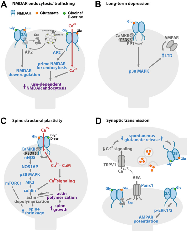
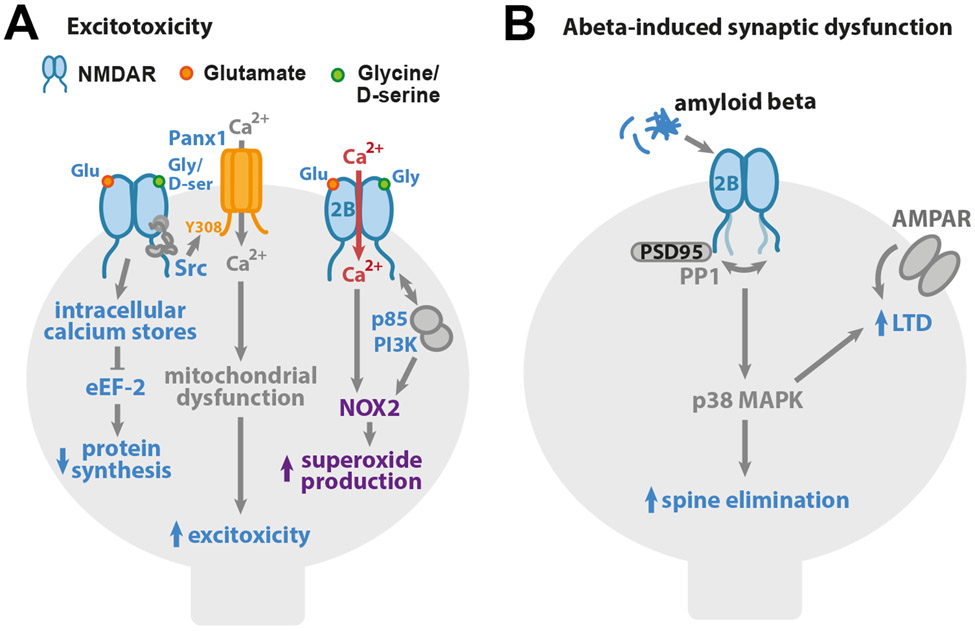
NMDA receptors play vital roles in a broad array of essential brain functions, from synaptic transmission and plasticity to learning and memory. Historically, the fundamental roles of NMDARs were attributed to their specialized properties of ion flux. More recently, it has become clear that NMDARs also signal in an ion flux-independent manner. Here, we review these non-ionotropic NMDAR signaling mechanisms that have been reported to contribute to a broad array of neuronal functions and dysfunctions including synaptic transmission and plasticity, cell death and survival, and synaptic alterations associated with neurological disorders.

Keywords: Alzheimer's disease; Dendritic spine; Ion flux-independent; LTD; LTP; NMDA receptor; Non-ionotropic; Schizophrenia.


Conflict of interest statement

Declaration of interests: The authors declare no competing interests.

Figures

Figure 1.
Figure 1.. Non-ionotropic NMDAR signaling in neuronal physiology
(A) Binding of one or both co-agonists drives NMDAR downregulation. On the left, binding of NMDAR agonist and co-agonist drives dephosphorylation of GluN1 Y837 and GluN2A Y842, even when NMDAR ion flux is inhibited, leading to downregulation of the number of functional NMDARs. On the right, high concentrations of glycine (>10 μM) increase the interaction with AP2 and prime the NMDAR for dynamin-dependent endocytosis. (B) Glutamate binding to the NMDAR is sufficient to drive long-term depression (LTD) of synaptic strength, independent of ion flux through the NMDAR. p38 MAPK activation is required for LTD induction. Agonist binding to the NMDAR initiates a conformational change which drives the GluN1 C-tails apart and alters its interactions with CaMKII and PP1. (C) Ion flux-independent NMDAR signaling contributes to bidirectional spine structural plasticity. Glutamate binding with little or no ion flux through the NMDAR drives dendritic spine shrinkage via a signaling pathway that includes the interaction of nNOS and NOS1AP, and the activities of nNOS, p38 MAPK, CaMKII, MK2, and cofilin. Furthermore, the activity of mTORC1 supports a requirement for new protein synthesis. Notably, when this non-ionotropic NMDAR signaling pathway is activated in combination with strong calcium influx, it instead drives dendritic spine growth. (D) Non-ionotropic NMDAR signaling plays a role in the regulation of neurotransmitter release and synaptic strength. On the left, ligand binding to postsynaptic NMDARs activates Pannexin-1 channels through Src. Opening of Pannexin-1 drives clearance of AEA, a ligand for presynaptic TRPV1, from the synaptic cleft. With decreased AEA, TRPV1 closes, reducing calcium available for spontaneous release of glutamate. On the right bottom, in a second mechanism, co-agonist binding to GluN2A-containing postsynaptic NMDARs activates ERK1/2 and increases synaptic strength. On the right top, activation of presynaptic NMDARs by agonist, or both agonist and co-agonist, enhances spontaneous release of glutamate in an ion flux-independent manner.
Figure 2.
Figure 2.. Non-ionotropic NMDAR signaling in neuronal dysfunction and disease
(A) Non-ionotropic NMDAR signaling contributes to excitotoxicity. Ligand binding to the NMDAR drives opening of intracellular calcium stores and inhibition of eEF-2 and protein synthesis. Glutamate and co-agonist binding to the NMDAR leads to Src kinase activation and the opening of Pannexin-1 channels and Ca2+ influx, which drives the mitochondrial dysfunction, contributing to excitotoxicity. Ligand binding to GluN2B-containing NMDARs increases binding of p85 to GluN2B C-tail, removing the PI3K regulatory domain so PI3K can activate NOX2 and superoxide production that results in excitotoxicity. (B) Synaptic dysfunction associated with Alzheimer’s disease can be mediated through non-ionotropic NMDAR signaling, β-amyloid acting via GluN2B subunit-containing NMDAR complexes results in activation of p38 MAPK and drives LTD and dendritic spine elimination.

References

    1. Aarts M, Liu Y, Liu L, Besshoh S, Arundine M, Gurd JW, Wang YT, Salter MW, and Tymianski M (2002). Treatment of ischemic brain damage by perturbing NMDA receptor- PSD-95 protein interactions. Science 298, 846–850. - PubMed
    1. Abrahamsson T, Chou CYC, Li SY, Mancino A, Costa RP, Brock JA, Nuro E, Buchanan KA, Elgar D, Blackman AV, et al. (2017). Differential Regulation of Evoked and Spontaneous Release by Presynaptic NMDA Receptors. Neuron 96, 839–855 e835. - PubMed
    1. Aow J, Dore K, and Malinow R (2015). Conformational signaling required for synaptic plasticity by the NMDA receptor complex. Proc Natl Acad Sci U S A 112, 14711–14716. - PMC - PubMed
    1. Babiec WE, Guglietta R, Jami SA, Morishita W, Malenka RC, and O'Dell TJ (2014). Ionotropic NMDA receptor signaling is required for the induction of long-term depression in the mouse hippocampal CA1 region. J Neurosci 34, 5285–5290. - PMC - PubMed
    1. Bai D, Muller RU, and Roder JC (2002). Non-ionotropic cross-talk between AMPA and NMDA receptors in rodent hippocampal neurones. J Physiol 543, 23–33. - PMC - PubMed

Publication types

MeSH terms

Substances