. 2022 May;298(5):101818.
doi: 10.1016/j.jbc.2022.101818. Epub 2022 Mar 10.

Gonadal white adipose tissue is important for gametogenesis in mice through maintenance of local metabolic and immune niches

Affiliations

Gonadal white adipose tissue is important for gametogenesis in mice through maintenance of local metabolic and immune niches

Chao-Fan Yang et al. J Biol Chem. 2022 May.

Abstract

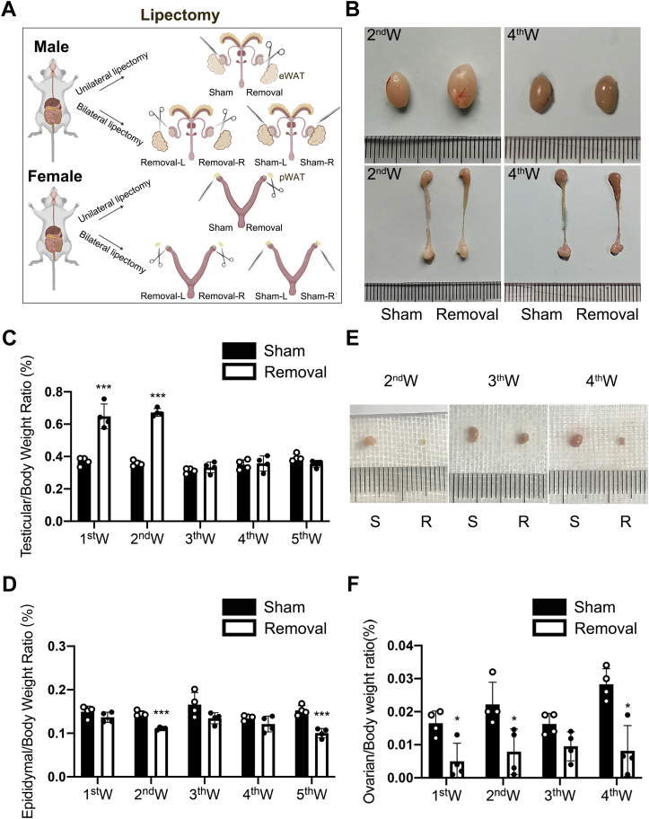
Gonadal white adipose tissue (gWAT) can regulate gametogenesis via modulation of neuroendocrine signaling. However, the effect of gWAT on the local microenvironment of the gonad was largely unknown. Herein, we ruled out that gWAT had a neuroendocrine effect on gonad function through a unilateral lipectomy strategy, in which cutting off epididymal white adipose tissue could reduce seminiferous tubule thickness and decrease sperm counts only in the adjacent testis and epididymis of the affected gonad. Consistent with the results in males, in females, ovary mass was similarly decreased by lipectomy. We determined that the defects in spermatogenesis were mainly caused by augmented apoptosis and decreased proliferation of germ cells. Transcriptome analysis suggested that lipectomy could disrupt immune privilege and activate immune responses in both the testis and ovary on the side of the lipectomy. In addition, lipidomics analysis in the testis showed that the levels of lipid metabolites such as free carnitine were elevated, whereas the levels of glycerophospholipids such as phosphatidylcholines and phosphatidylethanolamines were decreased, which indicated that the metabolic niche was also altered. Finally, we show that supplementation of phosphatidylcholine and phosphatidylethanolamine could partially rescue the observed phenotype. Collectively, our findings suggest that gWAT is important for gonad function by not only affecting whole-body homeostasis but also via maintaining local metabolic and immune niches.

Keywords: gWAT; gametogenesis; immune niche; lipectomy; metabolic niche.


Conflict of interest statement

Conflict of interest The authors declare that they have no conflicts of interest with the contents of this article.

Figures

Figure 1
Figure 1
Lipectomy leads to the reduction of ovarian mass and epididymal mass, whereas testis mass first increases and then decreases.A, unilateral lipectomy and bilateral lipectomy of gWAT. Unilateral lipectomy refers to the sham surgery on the left, whereas it refers to the removal surgery on the right in the same mice. Bilateral lipectomy including bilateral removal surgery and bilateral sham surgery. B, representative images of testes and epididymis in 8-week-old mice at the second and fourth weeks after unilateral lipectomy. C, analysis of the testicular weight versus body weight ratio between the sham side and the removal side at first week to fifth week after unilateral lipectomy in 8-week-old mice, n = 4. D, analysis of the epididymal weight versus body weight ratio between the sham side and the removal side at first week to fifth week after unilateral lipectomy in 8-week-old mice, n = 4. E, representative images of ovaries in 5-week-old mice with unilateral removal surgery at the second week to the fourth week. F, analysis of the ovarian weight versus body weight ratio at the first week to the fourth week after unilateral lipectomy in 5-week-old mice, n = 4. ∗p < 0.05, ∗∗∗p < 0.001. gWAT, gonadal white adipose tissue.
Figure 2
Figure 2
The seminiferous tubule cellular lining thickness, sperm counts, and sperm quality were significantly decreased in mice that underwent epididymal adipose lipectomy.A, H&E staining of testis of the sham side and the removal side at the fourth week after partial unilateral lipectomy in 8-week-old mice; 1/3, remove a third of the eWAT; 2/3, remove two-thirds of the eWAT. B, H&E staining of the epididymis of the sham side and removal side at the fourth week after unilateral lipectomy in 8-week-old mice. C, representative photographs of the sperm from the sham side and removal side at the fourth week after unilateral lipectomy in 8-week-old mice. D, sperm motility analysis of the sham side and removal side at the fourth week after unilateral lipectomy in 8-week-old mice by CASA analysis, n = 7. E, sperm count analysis of the sham side and removal side at the fourth week after unilateral lipectomy in 8-week-old mice, n = 5. F, sperm PR analysis of  the sham side and removal side at the fourth week after unilateral lipectomy in 8-week-old mice by CASA analysis , n = 6. G, analysis of the abnormal sperm rate of the sham side and removal side at the fourth week after unilateral lipectomy in 8-week-old mice, n = 5. ∗∗p < 0.01, ∗∗∗p < 0.001, and ∗∗∗∗p < 0.0001. CASA, computer-assisted semen analysis; eWAT, epididymal white adipose tissue; PR, progressive movement.
Figure 3
Figure 3
Periovarian adipose tissue is important to follicle development, but it does not work by affecting hormone levels.A, H&E staining of ovaries of the sham side and removal side at the fourth weeks after unilateral lipectomy in 5-week-old mice. B, analysis of the number of follicles at different stages at the fourth week after unilateral lipectomy in 5-week-old mice, ∗p < 0.05, ∗∗p < 0.01, and ∗∗∗p < 0.001, n = 5. C, vaginal smear assays were conducted daily to monitor estrous cycles after lipectomy and sham surgery in 5-week-old mice. The graph shows representative cycles from the two groups. Each box indicates one animal, and dots represent a day; n = 5. D and E, analysis of serum concentrations of FSH and LH lever as measured by RIA in 5-week-old female mice after pWAT_ unilateral lipectomy and sham surgery, n = 7. F, analysis of serum concentrations of T lever as measured by RIA in 5-week-old female mice after pWAT unilateral lipectomy and sham surgery, n = 7. G, analysis of serum concentrations of E2 lever as measured by RIA in 5-week-old female mice after pWAT unilateral lipectomy and sham surgery, n = 7. D, diestrus; E, estrus; E2, estradiol 2; FSH, follicle-stimulating hormone; LH, luteinizing hormone; M, metestrus; P, proestrus; pWAT, periovarian white adipose tissue; RIA, radioimmunoassay; T, testosterone.
Figure 4
Figure 4
Epididymal adipose lipectomy mainly results in loss of spermatids and increased apoptosis of germ cells in seminiferous tubules.A, immunohistochemistry staining of proliferating marker PHH3 in testes of the sham side and removal side at the first week and fourth week after unilateral lipectomy. B, analysis of PHH3-positive cell number in testes of the sham side and removal side at the first week and fourth week after unilateral lipectomy, n = 5. C, TUNEL apoptosis detection of cell in testes of the sham side and removal side. D, analysis of TUNEL-positive cell number in testes of the sham side and removal side at the first week and fourth week after unilateral lipectomy, n = 5. E, immunofluorescence staining of the SC marker WT1 in testes of the sham side and removal side at the fourth week after unilateral lipectomy. F, immunofluorescence staining of the GC marker mouse vasa homolog (MVG) in testes of the sham side and removal side. G, immunofluorescence staining of the spermatogonia stem cells marker Plzf in testes of the sham side and removal side at the fourth week after unilateral lipectomy. H, immunofluorescence staining of the spermatocyte marker Sycp3 in testes of the sham side and removal side at the fourth week after unilateral lipectomy. I, PAS staining of acrosome. PAS-positive substances (glycogen or polysaccharide) are red. Red arrow points to the round spermatids. J, analysis of round spermatid number in testes of the sham side and removal side at the fourth week after unilateral lipectomy, n = 5. ∗p < 0.05, ∗∗p < 0.01, and ∗∗∗p < 0.001. GC, germ cell; PAS, periodic acid–Schiff; PHH3, phosphohistone H3; SC, Sertoli cell.
Figure 5
Figure 5
Unilateral lipectomy damages the immune and metabolic local microenvironment of testes and ovaries.A, volcano plot showing the number of transcripts with upregulated and downregulated expression between the sham and removal groups in testes. Log2 fold change ≥1.2. B, results of KEGG enrichment analysis of the genes with upregulated and downregulated expression between the sham and removal groups in testes. p < 0.05. C, results of GO enrichment analysis of the genes with upregulated and downregulated expression between the sham and removal groups in testes. p < 0.05. D, volcano plot showing the number of transcripts with upregulated and downregulated expression between the sham and removal groups in ovaries. Log2 fold change ≥1.2. E, results of KEGG enrichment analysis of the genes with upregulated and downregulated expression between the sham and removal groups in ovaries. p < 0.05. F, results of GO enrichment analysis of the genes with upregulated and downregulated expression between the sham and removal groups in ovaries. p < 0.05. GO, Gene Ontology; KEGG, Kyoto Encyclopedia of Genes and Genomes.
Figure 6
Figure 6
Unilateral lipectomy altered immune environment and BTB integrity but not because of nonspecific damage.A and B, verification experiment of qPCR analysis of eight differentially expressed genes between the sham and removal groups. n = 6. C, the expression of inflammation-related genes in the testis at the second week after lipectomy by RT–qPCR analysis. n = 6. D, the expression of inflammation-related genes in the testis at the fourth week after lipectomy by RT–qPCR analysis. n = 6. E, immunofluorescence staining of the mouse major macrophage marker F4/80 in testes from the sham side and removal side at the fourth week after unilateral lipectomy. F, immunohistochemistry staining of the mouse major macrophage marker CD11b in testes from the sham side and removal side at the fourth week after unilateral lipectomy. G, immunohistochemistry staining of the inflammatory factor IL6 in testes from the sham side and removal side at the fourth week after unilateral lipectomy. H, immunohistochemistry staining of the inflammatory factor TNF-α in testes from the sham side and removal side at the fourth week after unilateral lipectomy. I, immunofluorescence staining of the BTB structure marker ZO-1 in testes from the sham side and removal side at the fourth week after unilateral lipectomy in 8-week-old mice. J, fluorescence distribution in sham and removal mice testis after biotin injection in 8-week-old mice. The asterisk indicates the fluorescence penetrating into seminiferous tubules. ∗p < 0.05, ∗∗p < 0.01, and ∗∗∗p < 0.001. BTB, blood–testis barrier; IL6, interleukin 6; qPCR, quantitative PCR; TNF-α, tumor necrosis factor-α.
Figure 7
Figure 7
Unilateral lipectomy alters the metabolic niche.A, PCA showing the distinction between the sham group and removal group. n = 5. B, volcano plot showing the increased and decreased lipid metabolites between the sham and removal groups. Fold change ≥1. C, details of 10 different lipid metabolites. D, verification of ELISA analysis of free carnitine, n = 6. E, verification of ELISA analysis of PC and PE, n ≥ 5. F, H&E staining of testis from 8-week-old lipectomy mice of PC + PE diet and normal diet at the fourth week after unilateral lipectomy. G, sperm count analysis of the sperm from 8-week-aged lipectomy mice of PC + PE diet and normal diet at the fourth week after unilateral lipectomy, n = 5. H, immunofluorescence staining of the GC marker MVH in testes from 8-week-aged lipectomy mice of PC + PE diet and normal diet at the fourth week after unilateral lipectomy. I, statistical analysis of MVH-positive cells, n = 4. ∗p < 0.05. GC, germ cell; MVH, mouse vasa homolog; PC, phosphatidylcholine; PCA, principal component analysis; PE, phosphatidylethanolamine.

References

    1. Du Plessis S.S., Cabler S., McAlister D.A., Sabanegh E., Agarwal A. The effect of obesity on sperm disorders and male infertility. Nat. Rev. Urol. 2010;7:153–161. - PubMed
    1. Pasquali R., Pelusi C., Genghini S., Cacciari M., Gambineri A. Obesity and reproductive disorders in women. Hum. Reprod. Update. 2003;9:359–372. - PubMed
    1. Hohos N.M., Skaznik-Wikiel M.E. High-fat diet and female fertility. Endocrinology. 2017;158:2407–2419. - PMC - PubMed
    1. Yen S.S. Female hypogonadotropic hypogonadism. Hypothalamic amenorrhea syndrome. Endocrinol. Metab. Clin. North Am. 1993;22:29–58. - PubMed
    1. Starkey T.A., Lee R.A. Menstruation and fertility in anorexia nervosa. Am. J. Obstet. Gynecol. 1969;105:374–379. - PubMed

Publication types

LinkOut - more resources