. 2022 May:149:112802.
doi: 10.1016/j.biopha.2022.112802. Epub 2022 Mar 9.

Potential inhibitor for blocking binding between ACE2 and SARS-CoV-2 spike protein with mutations

Affiliations

Potential inhibitor for blocking binding between ACE2 and SARS-CoV-2 spike protein with mutations

Ming-Shao Tsai et al. Biomed Pharmacother. 2022 May.

Abstract

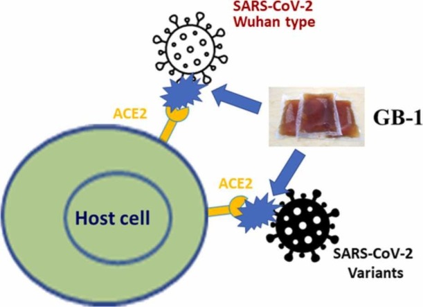
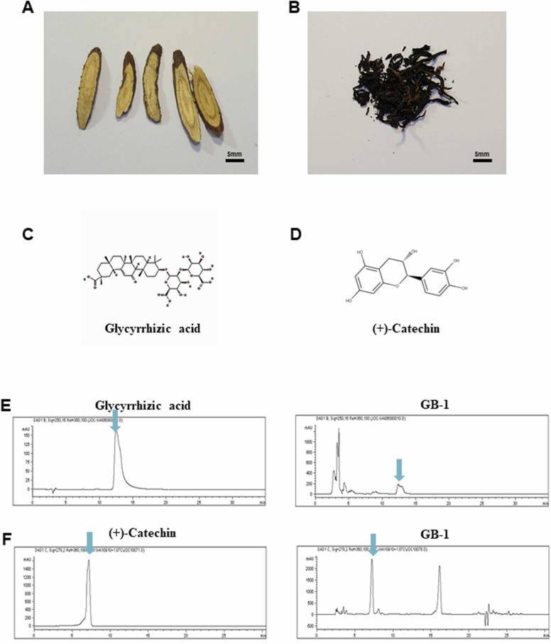
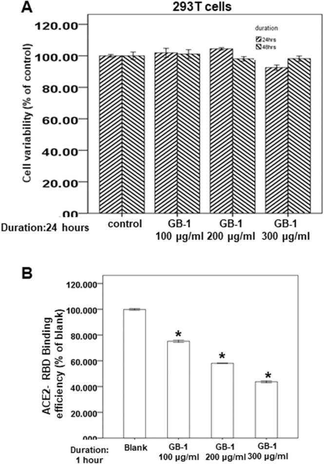
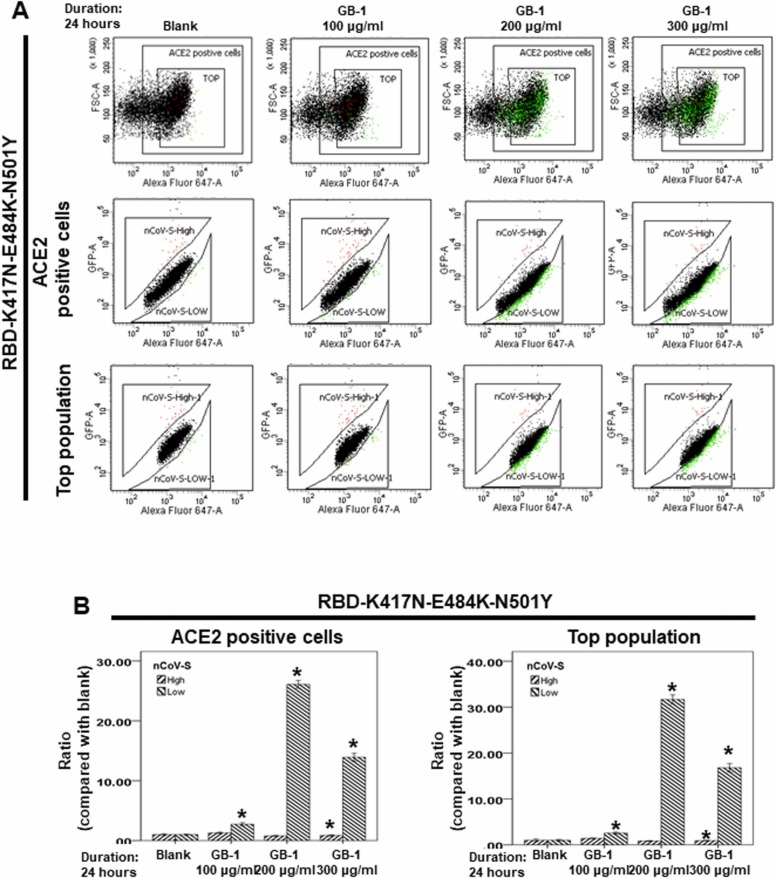
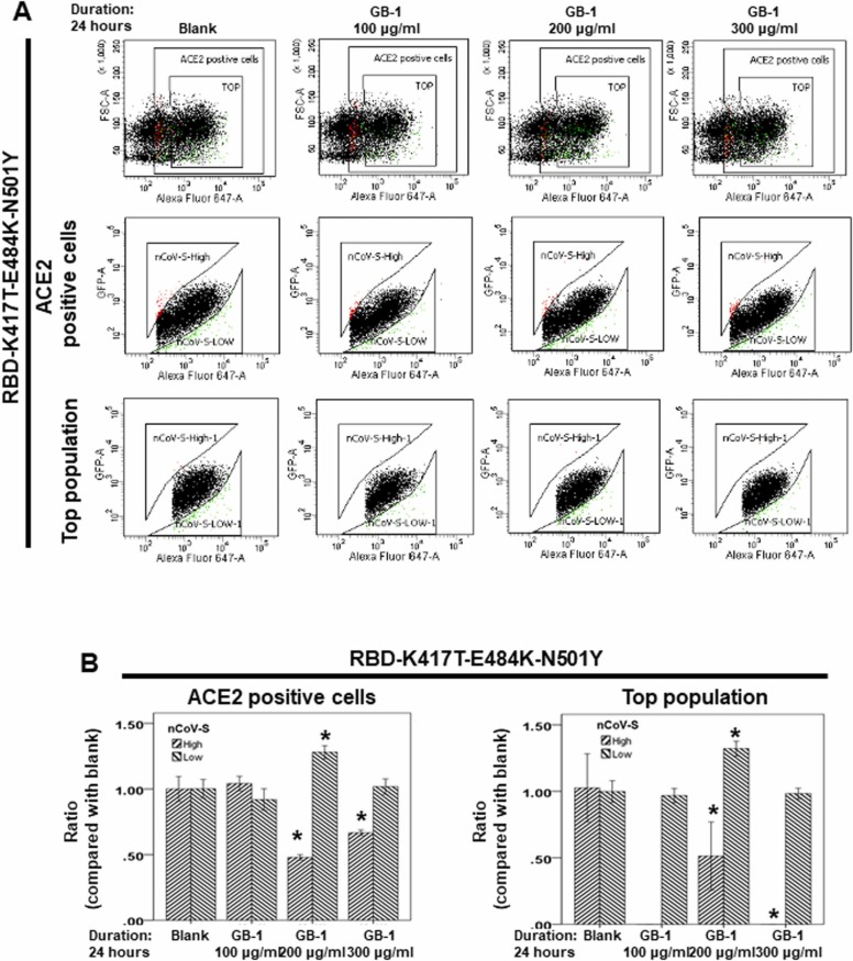
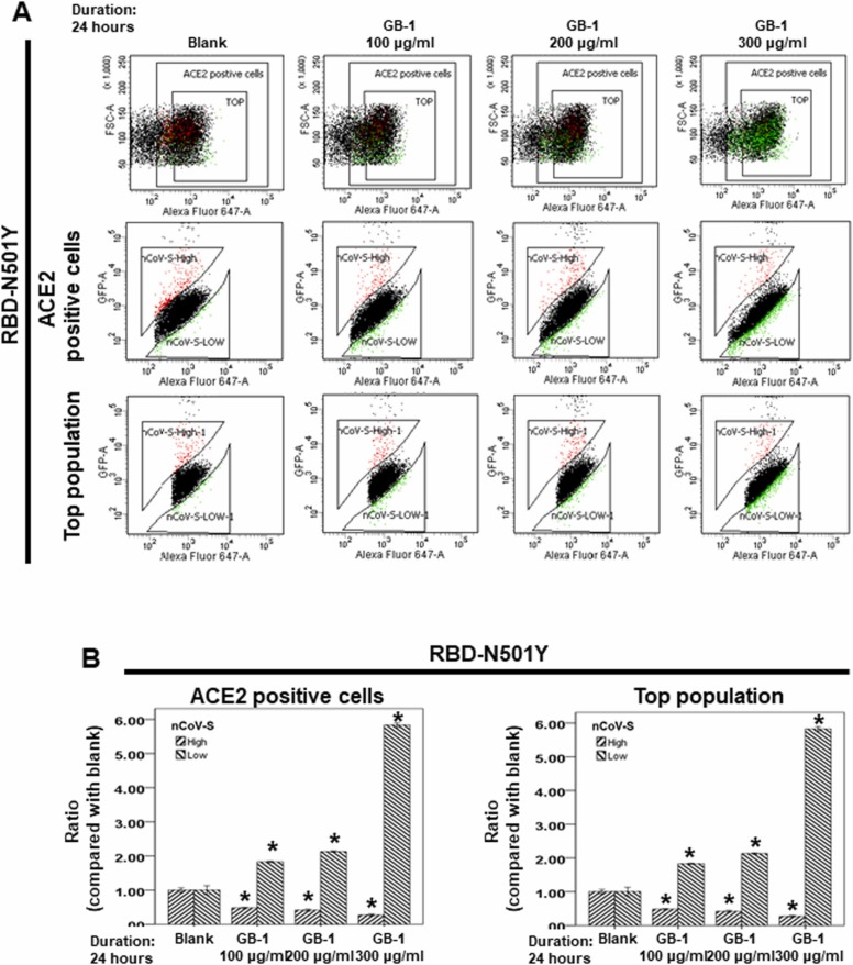
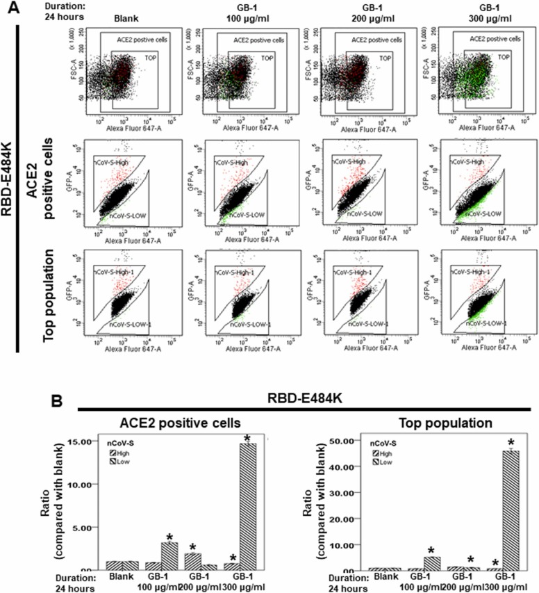
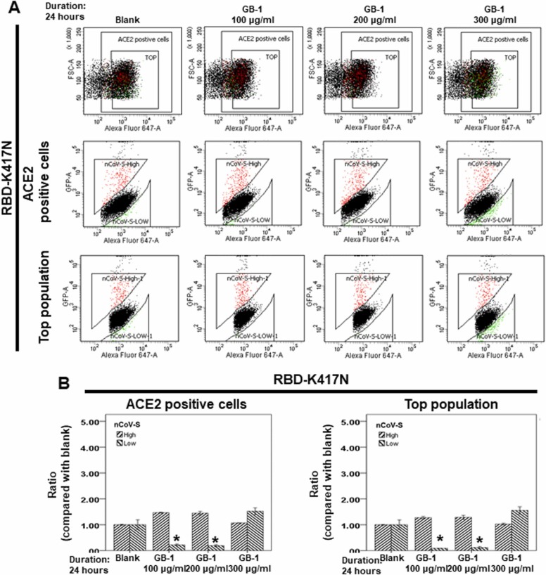
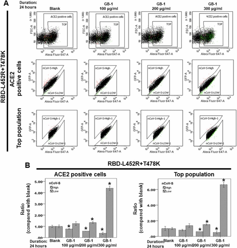
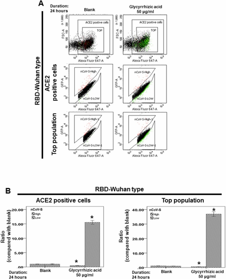
At the time of writing, more than 440 million confirmed coronavirus disease 2019 (COVID-19) cases and more than 5.97 million COVID-19 deaths worldwide have been reported by the World Health Organization since the start of the outbreak of the pandemic in Wuhan, China. During the COVID-19 pandemic, many variants of SARS-CoV-2 have arisen because of high mutation rates. N501Y, E484K, K417N, K417T, L452R and T478K in the receptor binding domain (RBD) region may increase the infectivity in several variants of SARS-CoV-2. In this study, we discovered that GB-1, developed from Chiehyuan herbal formula which obtained from Tian Shang Sheng Mu of Chiayi Puzi Peitian Temple, can inhibit the binding between ACE2 and RBD with Wuhan type, K417N-E484K-N501Y and L452R-T478K mutation. In addition, GB-1 inhibited the binding between ACE2 and RBD with a single mutation (E484K or N501Y), except the K417N mutation. In the compositions of GB-1, glycyrrhizic acid can inhibit the binding between ACE2 and RBD with Wuhan type, except K417N-E484K-N501Y mutation. Our results suggest that GB-1 could be a potential candidate for the prophylaxis of different variants of SARS-CoV-2 infection because of its inhibition of binding between ACE2 and RBD with different mutations (L452R-T478K, K417N-E484K-N501Y, N501Y or E484K).

Keywords: (+)-Catechin (Pubchem CID: 9064); COVID-19; GB-1; Glycyrrhizic acid; Glycyrrhizic acid (Pubchem CID: 14982); SARS-CoV-2; Spike protein.


Conflict of interest statement

The authors declare no conflict of interest.

Figures

ga1
Graphical abstract
Fig. 1
Fig. 1
Identification of reference compounds in GB-1 by HPLC analysis. (A) The photo of the slices of dry root of Glycyrrhiza uralensis. (B) The photo of the dry leaves of Camellia sinensis. (C) The structure of glycyrrhizic acid. (D) The structure of (+)-catechin. (E) HPLC chromatograms of glycyrrhizic acid and GB-1. (F) HPLC chromatograms of (+)-catechin and GB-1.
Fig. 2
Fig. 2
Effect of GB-1 on cell variability of the 293 T cell line and the interaction between ACE2 and RBD with Wuhan type. (A) 293 T cells were measured by XTT assay after indicated hours of culturing in the presence of GB-1. (B) The indicated compounds were tested to evaluate their ability to inhibit the binding of spike protein to immobilized ACE2 by the ACE2/SARS-CoV-2 spike inhibitor screening assay. All the results are representative of at least three independent experiments. (Error bars=mean±S.E.M. Asterisks (*) mark samples significantly different from control group with p < 0.05).
Fig. 3
Fig. 3
Effect of GB-1 on interaction between ACE2 and RBD with K417N-E484K-N501Y mutation. (A) Flow cytometry analysis of ACE2-Spike protein binding. 293 T cells with pCEP4-MYC-ACE2 plasmid were incubated with RBD (K417N-E484K-N501Y)-sfGFP-containing medium and co-stained with fluorescent anti-MYC Alexa 647 to detect surface ACE2 by flow cytometry. During analysis, the top population were chosen from the ACE2-positive population. Then, two subsets of the ACE2-positive population were collected: the top population (nCoV-S-High sort, red gate) and the bottom population (nCoV-S-Low sort, green gate) based on the fluorescence of bound RBD(K417N-E484K-N501Y)-sfGFP relative to ACE2 surface expression. (B) Quantitative results of nCoV-S-High sort and nCoV-S-low sort, which were presented as ratio compared with blank, in the top population or ACE2-positive population. All the results are representative of at least three independent experiments. (Error bars = mean±S.E.M. Asterisks (*) mark samples significantly different from control group with p < 0.05).
Fig. 4
Fig. 4
Effect of GB-1 on interaction between ACE2 and RBD with K417T-E484K-N501Y mutation. (A) Flow cytometry analysis of ACE2-Spike protein binding. 293 T cells with pCEP4-MYC-ACE2 plasmid were incubated with RBD (K417T-E484K-N501Y)-sfGFP-containing medium and co-stained with fluorescent anti-MYC Alexa 647 to detect surface ACE2 by flow cytometry. During analysis, the top population were chosen from the ACE2-positive population. Then, two subsets of the ACE2-positive population were collected: the top population (nCoV-S-High sort, red gate) and the bottom population (nCoV-S-Low sort, green gate) based on the fluorescence of bound RBD(K417T-E484K-N501Y)-sfGFP relative to ACE2 surface expression. (B) Quantitative results of nCoV-S-High sort and nCoV-S-low sort, which were presented as ratio compared with blank, in the top population or ACE2-positive population. All the results are representative of at least three independent experiments. (Error bars=mean±S.E.M. Asterisks (*) mark samples significantly different from control group with p < 0.05).
Fig. 5
Fig. 5
Effect of GB-1 on interaction between ACE2 and RBD with N501Y mutation. Flow cytometry analysis of ACE2-Spike protein binding. (A) 293 T cells with pCEP4-myc-ACE2 plasmid were incubated with RBD (N501Y)-sfGFP-containing medium and co-stained with fluorescent anti-MYC Alexa 647 to detect surface ACE2 by flow cytometry. During analysis, the top population were chosen from the ACE2-positive population. Then, two subsets of the ACE2-positive population were collected: the top population (nCoV-S-High sort, red gate) and the bottom population (nCoV-S-Low sort, green gate) based on the fluorescence of bound RBD (N501Y)-sfGFP relative to ACE2 surface expression. (B) Quantitative results of nCoV-S-High sort and nCoV-S-low sort, which were presented as ratio compared with blank, in the top population or ACE2-positive population. All the results are representative of at least three independent experiments. (Error bars=mean±S.E.M. Asterisks (*) mark samples significantly different from control group with p < 0.05).
Fig. 6
Fig. 6
Effect of GB-1 on interaction between ACE2 and RBD with E484K mutation. Flow cytometry analysis of ACE2-Spike protein binding. (A) 293 T cells with pCEP4-myc-ACE2 plasmid were incubated with RBD (E484K)-sfGFP-containing medium and co-stained with fluorescent anti-MYC Alexa 647 to detect surface ACE2 by flow cytometry. During analysis, the top population were chosen from the ACE2-positive population. Then, two subsets of the ACE2-positive population were collected: the top population (nCoV-S-High sort, red gate) and the bottom population (nCoV-S-Low sort, green gate) based on the fluorescence of bound RBD (E484K)-sfGFP relative to ACE2 surface expression. (B) Quantitative results of nCoV-S-High sort and nCoV-S-low sort, which were presented as ratio compared with blank, in the top population or ACE2-positive population. All the results are representative of at least three independent experiments. (Error bars=mean±S.E.M. Asterisks (*) mark samples significantly different from control group with p < 0.05).
Fig. 7
Fig. 7
Effect of GB-1 on interaction between ACE2 and RBD with K417N. Flow cytometry analysis of ACE2-Spike protein binding. (A) 293 T cells with pCEP4-myc-ACE2 plasmid were incubated with RBD (K417N)-sfGFP-containing medium and co-stained with fluorescent anti-MYC Alexa 647 to detect surface ACE2 by flow cytometry. During analysis, the top population were chosen from the ACE2-positive population. Then, two subsets of the ACE2-positive population were collected: the top population (nCoV-S-High sort, red gate) and the bottom population (nCoV-S-Low sort, green gate) based on the fluorescence of bound RBD (K417N)-sfGFP relative to ACE2 surface expression. (B) Quantitative results of nCoV-S-High sort and nCoV-S-low sort, which were presented as ratio compared with blank, in the top population or ACE2-positive population. All the results are representative of at least three independent experiments. (Error bars=mean±S.E.M. Asterisks (*) mark samples significantly different from control group with p < 0.05).
Fig. 8
Fig. 8
Effect of GB-1 on interaction between ACE2 and RBD with L452R-T478K mutation. (A) Flow cytometry analysis of ACE2-Spike protein binding. 293 T cells with pCEP4-MYC-ACE2 plasmid were incubated with RBD (L452R-T478K)-sfGFP-containing medium and co-stained with fluorescent anti-MYC Alexa 647 to detect surface ACE2 by flow cytometry. During analysis, the top population were chosen from the ACE2-positive population. Then, two subsets of the ACE2-positive population were collected: the top population (nCoV-S-High sort, red gate) and the bottom population (nCoV-S-Low sort, green gate) based on the fluorescence of bound RBD(L452R-T478K)-sfGFP relative to ACE2 surface expression. (B) Quantitative results of nCoV-S-High sort and nCoV-S-low sort, which were presented as ratio compared with blank, in the top population or ACE2-positive population. All the results are representative of at least three independent experiments. (Error bars=mean±S.E.M. Asterisks (*) mark samples significantly different from control group with p < 0.05).
Fig. 9
Fig. 9
Effect of glycyrrhizic acid on interaction between ACE2 and RBD with Wuhan type. (A) Flow cytometry analysis of ACE2-Spike protein binding. 293 T cells with pCEP4-MYC-ACE2 plasmid were incubated with RBD (Wuhan type)-sfGFP-containing medium and co-stained with fluorescent anti-MYC Alexa 647 to detect surface ACE2 by flow cytometry. During analysis, the top population were chosen from the ACE2-positive population. Then, two subsets of the ACE2-positive population were collected: the top population (nCoV-S-High sort, red gate) and the bottom population (nCoV-S-Low sort, green gate) based on the fluorescence of bound RBD (Wuhan type)-sfGFP relative to ACE2 surface expression. (B) Quantitative results of nCoV-S-High sort and nCoV-S-low sort, which were presented as ratio compared with blank, in the top population or ACE2-positive population. All the results are representative of at least three independent experiments. (Error bars=mean±S.E.M. Asterisks (*) mark samples significantly different from control group with p < 0.05).
Fig. 10
Fig. 10
Effect of glycyrrhizic acid on interaction between ACE2 and RBD with K417N-E484K-N501Y mutation. (A) Flow cytometry analysis of ACE2-Spike protein binding. 293 T cells with pCEP4-MYC-ACE2 plasmid were incubated with RBD (K417N-E484K-N501Y)-sfGFP-containing medium and co-stained with fluorescent anti-MYC Alexa 647 to detect surface ACE2 by flow cytometry. During analysis, the top population were chosen from the ACE2-positive population. Then, two subsets of the ACE2-positive population were collected: the top population (nCoV-S-High sort, red gate) and the bottom population (nCoV-S-Low sort, green gate) based on the fluorescence of bound RBD (K417N-E484K-N501Y)-sfGFP relative to ACE2 surface expression. (B) Quantitative results of nCoV-S-High sort and nCoV-S-low sort, which were presented as ratio compared with blank, in the top population or ACE2-positive population. All the results are representative of at least three independent experiments. (Error bars=mean±S.E.M. Asterisks (*) mark samples significantly different from control group with p < 0.05).

Similar articles

Cited by

References

    1. Walls A.C., Park Y.J., Tortorici M.A., Wall A., McGuire A.T., Veesler D. Structure, function, and antigenicity of the SARS-CoV-2 spike glycoprotein. Cell. 2020 - PMC - PubMed
    1. Yan R., Zhang Y., Li Y., Xia L., Guo Y., Zhou Q. Structural basis for the recognition of the SARS-CoV-2 by full-length human ACE2. Science. 2020 - PMC - PubMed
    1. Hoffmann M., Kleine-Weber H., Schroeder S., Kruger N., Herrler T., Erichsen S., Schiergens T.S., Herrler G., Wu N.H., Nitsche A., Muller M.A., Drosten C., Pohlmann S. SARS-CoV-2 cell entry depends on ACE2 and TMPRSS2 and is blocked by a clinically proven protease inhibitor. Cell. 2020 - PMC - PubMed
    1. Duffy S. Why are RNA virus mutation rates so damn high? PLoS Biol. 2018;16(8) - PMC - PubMed
    1. Li Q., Wu J., Nie J., Zhang L., Hao H., Liu S., Zhao C., Zhang Q., Liu H., Nie L., Qin H., Wang M., Lu Q., Li X., Sun Q., Liu J., Zhang L., Li X., Huang W., Wang Y. The impact of mutations in SARS-CoV-2 Spike on viral infectivity and antigenicity. Cell. 2020;182(5):1284–1294. e9. - PMC - PubMed

Substances