. 2022 Mar 12;12(1):4321.
doi: 10.1038/s41598-022-08052-z.

The distinctive roles played by the superoxide dismutases of the extremophile Acinetobacter sp. Ver3

Affiliations

The distinctive roles played by the superoxide dismutases of the extremophile Acinetobacter sp. Ver3

Bruno Alejandro Steimbrüch et al. Sci Rep. .

Abstract

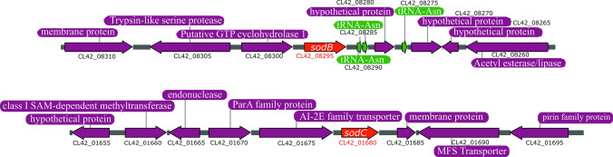
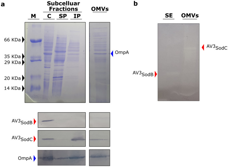
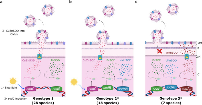
Acinetobacter sp. Ver3 is a polyextremophilic strain characterized by a high tolerance to radiation and pro-oxidants. The Ver3 genome comprises the sodB and sodC genes encoding an iron (AV3SodB) and a copper/zinc superoxide dismutase (AV3SodC), respectively; however, the specific role(s) of these genes has remained elusive. We show that the expression of sodB remained unaltered in different oxidative stress conditions whereas sodC was up-regulated in the presence of blue light. Besides, we studied the changes in the in vitro activity of each SOD enzyme in response to diverse agents and solved the crystal structure of AV3SodB at 1.34 Å, one of the highest resolutions achieved for a SOD. Cell fractionation studies interestingly revealed that AV3SodB is located in the cytosol whereas AV3SodC is also found in the periplasm. Consistently, a bioinformatic analysis of the genomes of 53 Acinetobacter species pointed out the presence of at least one SOD type in each compartment, suggesting that these enzymes are separately required to cope with oxidative stress. Surprisingly, AV3SodC was found in an active state also in outer membrane vesicles, probably exerting a protective role. Overall, our multidisciplinary approach highlights the relevance of SOD enzymes when Acinetobacter spp. are confronted with oxidizing agents.


Conflict of interest statement

The authors declare no competing interests.

Figures

Figure 1
Figure 1
Environment of sod genes in Acinetobacter sp. Ver3. Schematic representation of the Acinetobacter sp. Ver3 sodB (up) and sodC (down) loci, present in contigs JFYL01000023 and JFYL01000003, respectively. sod genes are shown in red while proximal genes are displayed in purple. The locus tag is provided below each gene. Three tRNAs (green) are encoded in the sodB region.
Figure 2
Figure 2
Sequence alignment and phylogenetic analysis of SOD enzymes. (a) Sequence alignment of FeSOD enzymes from selected species. Amino acids highlighted in purple correspond to conserved metal ligands. The motifs AAQ and GGH (in MnSODs, the E. coli enzyme is shown for comparison) are shown in red and green, respectively; the motif DVWEHAYYID comprising metal binding residues is boxed. Secondary structure elements observed in the crystal structure of AV3SodB are depicted as red and green lines below the sequence alignment. Multiple sequence alignments were performed using MAFFT version 7.475 (https://mafft.cbrc.jp/alignment/software/) with default parameters. (b) Phylogenetic tree of Fe-MnSODs of known structure. A clade of MnSODs (red) and two of FeSODs (blue and green) are distinguished. Cambialistics Fe/MnSODs are in bold. (c) Sequence alignment of CuZnSOD enzymes from selected species. Conserved metal ligands are shown in colour: copper ligands in vermillion, zinc ligands in green and a histidine residue that coordinates both metal ions in blue. Predicted signal peptides are signalled in red and lipobox sequences are boxed. Secondary structure elements predicted by Jalview are depicted as red and green lines below the sequence alignment. (d) Phylogenetic tree built for CuZnSOD sequences retrieved from the NCBI or the PDB (light blue boxes). Note that the signal peptide was removed from each sequence prior to alignment. Trees were built based on the amino acid sequence alignments of the Acinetobacter sp. Ver3 SOD enzymes by using the Maximum likelihood method (with a WAG + I + G4 substitution model). The bioinformatic software IQ-TREE multicore version 1.6.11 was employed to generate both trees and their reliability was tested by bootstrapping with 10,000 repetitions. Results were visualized using the online toll iTOL V5.7 (https://itol.embl.de/).
Figure 3
Figure 3
Biochemical characterization of recombinant Acinetobacter sp. Ver3 SODs. AV3SodB (a) and AV3SodC-P (b) were analysed by SDS-PAGE and Coomassie Blue staining (left panels). SOD activity was revealed in situ in nondenaturing polyacrylamide gels (right panels). M: molecular weight marker. The SDS-PAGE and the polyacrylamide gels were cropped to create the figure and the full-length gels are presented in Supplementary Figure S5. (c) Thermostability of AV3SodB and (d) AV3SodC-P. In each case, the residual SOD activity was measured after different heat treatments. (e) pH tolerance of AV3SodB (blue line) and AV3SodC-P (red line). In each case, the residual SOD activity was measured at pH 7.8 after incubating the enzyme 1 h at 25ºC in buffers with different pH values. (f) Effect of diverse chemical agents on AV3SodB and AV3SodC-P activity. In each case, the residual SOD activity was measured after incubating the enzyme 1 h at 25º C with the compound. In all experiments, SOD activity was determined spectrophotometrically by inhibition of the xanthine/xanthine oxidase-induced reduction of cytochrome c, at 25º C; the activity of the untreated enzyme was defined as 100%. The reported values correspond to the mean of four measurements in three replicates of the experiment; bars indicate standard deviations.
Figure 4
Figure 4
Crystal structure of AV3SodB. (a) Although there is a single molecule of AV3SodB per asymmetric unit, two contiguous molecules (in beige and blue, the interface area between them is informed in Å2) related by crystallographic symmetry (oval symbol) give rise to a dimer of the protein which presents a quaternary structure similar to that observed for the E. coli FeSOD (PDB code 1ISA, in Gray, superimposed to AV3SodB) (on the left). Proteins are shown in ribbon representation. The surface of AV3SodB is depicted in gray. On the right, a monomer of AV3SodB is shown in rainbow colours, with helices represented as cylinders. Secondary structure elements conserved in FeSODs are indicated (see also Fig. 2a). NTD, N-terminal domain; CTD, C-terminal domain. An inset of the interface between AV3SodB molecules in a protein dimer is shown. Residues involved in intermolecular hydrogen bonds or salt bridges are depicted in stick representation. Iron ions are shown in this and subsequent panels as orange spheres. (b) Active site of AV3SodB. Metal ligands are represented as sticks. Interatomic distances are informed (in Å). One of the axial ligands is a water molecule (red sphere). (c) A conserved network of hydrogen bonds involving active site residues. Interatomic interactions are depicted as dashed lines. The mesh in panels (b) and (c) corresponds to the crystallographic 2mFo–DC electron density map contoured to 2.0 σ.
Figure 5
Figure 5
AV3SodB and AV3SodC subcellular localization. (A) Coomassie stained SDS-PAGE (upper panel) and Western Blot assay (lower panel) of cytosolic (C), soluble and insoluble periplasmic (SP and IP, respectively) fractions (7 µg of total proteins), and OMVs (15 µL from a 500X culture concentrate) obtained from Acinetobacter sp. Ver3. Specific antibodies raised against AV3SodB and AV3SodC (red arrows) were used. The detection of OmpA (blue arrow) with specific antibodies against the A. baumannii protein was used as a control; OmpA has been shown to be loaded into OMVs. (B) In-gel assessment of SOD activity present in a soluble extract (SE) (1.5 µg of total proteins) and OMVs (15 µL from a 500X culture concentrate) of Acinetobacter sp. Ver3. M: molecular weight marker. The specificity of the antibodies was tested showing that there is no cross-reactivity (Fig. S4). The SDS-PAGEs and the blots were cropped to create the figure. Full-length gels/blots are presented in Supplementary Figure S5.
Figure 6
Figure 6
sodB and sodC response to pro-oxidant challenges. (a) Relative levels of sodB and sodC transcription in untreated Acinetobacter sp. Ver3 cells (black bars) or after exposure to 1 mM H2O2 (red bars), 0.5 mM Methyl Viologen (MV) (green bars), 900 J.m-2 ultraviolet (UV) (yellow bars) or blue light (blue bars). The mean for the housekeeping genes recA and rpoB was used as a normalizer. Asterisks indicate significant differences among control and treated samples, as determined by analysis of variance (ANOVA) and Tukey’s multiple comparison test. In each case, the average value and the standard deviation of four biological replicates are shown. (b) Anti-AV3SodC immunoblot of a soluble extract (SE, 7 µg of total proteins) and OMVs (15 µL from a 500X culture concentrate) of Acinetobacter sp. Ver3 grown over 26 h in the presence of blue light (BL, 5 μmol.m-2.s-1). A control (C) culture was also included. The full-length blot is presented in Supplementary Figure S5.
Figure 7
Figure 7
Proposed model schematizing the subcellular localization of SODs encoded by Acinetobacter spp. according to their genotype. (a) Strains with a type 1 genotype harbour sodB and sodC genes, coding for a FeSOD and a CuZnSOD, which are located in the cytosolic (C) and periplasmic (P) spaces, respectively. The CuZnSOD could also be loaded into OMVs, as shown for Acinetobacter sp. Ver3 strain. (b) Strains with a type 2 genotype additionally harbour a sodAc gene, coding for a cytosolic MnSOD (cMnSOD). (c) Strains with a type 3 genotype bear sodB and sodAp genes, coding for a FeSOD and a periplasmic MnSOD (pMnSOD), respectively. In 12 out of the 19 strains in this group, a sodAc gene is also present. *One strain of A. beijerinckii is present in each of these groups. IM: inner membrane; OM: outer membrane. Created with BioRender.com.

References

    1. Albarracín VH, Gärtner W, Farias ME. Forged under the sun: Life and art of extremophiles from Andean Lakes. Photochem. Photobiol. 2016;92:14–28. - PubMed
    1. Ordoñez OF, Flores MR, Dib JR, Paz A, Farías ME. Extremophile culture collection from Andean Lakes: Extreme pristine environments that host a wide diversity of microorganisms with tolerance to UV radiation. Microb. Ecol. 2009;58:461–473. - PubMed
    1. Flores R, Ordoñez MF, Maldonado J, Farías E. Isolation of UV-B resistant bacteria from two high altitude Andean lakes (4,400 m) with saline and non saline conditions. J. Gen. Appl. Microbiol. 2009;55:447–458. - PubMed
    1. Albarracín VH, et al. Extremophilic Acinetobacter strains from high-altitude lakes in Argentinean Puna: Remarkable UV-B resistance and efficient DNA damage repair. Orig. Life Evol. Biosph. 2012;42:201–221. - PubMed
    1. Di Capua C, Bortolotti A, Farías ME, Cortez N. UV-resistant Acinetobacter sp. isolates from Andean wetlands display high catalase activity. FEMS Microbiol. Lett. 2011;317:181–189. - PubMed

Publication types

MeSH terms