

Neural oscillations during motor imagery of complex gait: an HdEEG study
- PMID: 35279682
- PMCID: PMC8918338
- DOI: 10.1038/s41598-022-07511-x
Neural oscillations during motor imagery of complex gait: an HdEEG study
Abstract
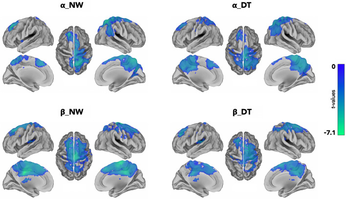
The aim of this study was to investigate differences between usual and complex gait motor imagery (MI) task in healthy subjects using high-density electroencephalography (hdEEG) with a MI protocol. We characterized the spatial distribution of α- and β-bands oscillations extracted from hdEEG signals recorded during MI of usual walking (UW) and walking by avoiding an obstacle (Dual-Task, DT). We applied a source localization algorithm to brain regions selected from a large cortical-subcortical network, and then we analyzed α and β bands Event-Related Desynchronizations (ERDs). Nineteen healthy subjects visually imagined walking on a path with (DT) and without (UW) obstacles. Results showed in both gait MI tasks, α- and β-band ERDs in a large cortical-subcortical network encompassing mostly frontal and parietal regions. In most of the regions, we found α- and β-band ERDs in the DT compared with the UW condition. Finally, in the β band, significant correlations emerged between ERDs and scores in imagery ability tests. Overall we detected MI gait-related α- and β-band oscillations in cortical and subcortical areas and significant differences between UW and DT MI conditions. A better understanding of gait neural correlates may lead to a better knowledge of pathophysiology of gait disturbances in neurological diseases.
© 2022. The Author(s).
Conflict of interest statement
The authors declare no competing interests.
Figures


References
-
- Avanzino L, Lagravinese G, Abbruzzese G, Pelosin E. Relationships between gait and emotion in Parkinson’s disease: A narrative review. Gait Posture. 2018;65:57–64. - PubMed
-
- Mirelman A, et al. Addition of a non-immersive virtual reality component to treadmill training to reduce fall risk in older adults (V-TIME): A randomised controlled trial. Lancet. 2016;388:1170–1182. - PubMed
-
- Fukuyama H, et al. Brain functional activity during gait in normal subjects: A SPECT study. Neurosci. Lett. 1997;228:183–186. - PubMed
Publication types
MeSH terms
Grants and funding
LinkOut - more resources
Full Text Sources
