Review
. 2022 Apr;127(4):369-382.
doi: 10.1007/s11547-022-01473-w. Epub 2022 Mar 13.

Extracorporeal membrane oxygenation (ECMO) in COVID-19 patients: a pocket guide for radiologists

Affiliations
Review

Extracorporeal membrane oxygenation (ECMO) in COVID-19 patients: a pocket guide for radiologists

Michela Gabelloni et al. Radiol Med. 2022 Apr.

Abstract

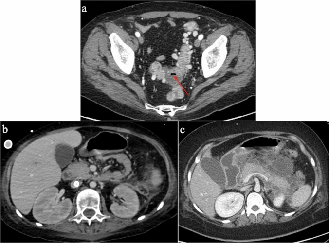
During the coronavirus disease 19 (COVID-19) pandemic, extracorporeal membrane oxygenation (ECMO) has been proposed as a possible therapy for COVID-19 patients with acute respiratory distress syndrome. This pictorial review is intended to provide radiologists with up-to-date information regarding different types of ECMO devices, correct placement of ECMO cannulae, and imaging features of potential complications and disease evolution in COVID-19 patients treated with ECMO, which is essential for a correct interpretation of diagnostic imaging, so as to guide proper patient management.

Keywords: Acute respiratory distress syndrome (ARDS); Computed tomography; Coronavirus disease 19 (COVID-19); Extracorporeal membrane oxygenation (ECMO); Severe acute respiratory syndrome coronavirus 2 (SARS-CoV-2); X-ray.


Conflict of interest statement

The authors declare that they have no competing interest.

Figures

Fig. 1
Fig. 1
Chest radiographs (antero-posterior view) showing a correct placement of VV-ECMO, b correct placement of femorofemoral ECMO, c correct placement of double lumen ECMO, d incorrect placement of femorofemoral ECMO. The tip of the drainage cannula (arrow) is too close to the tip of the return cannula
Fig. 2
Fig. 2
Chest radiographs a before and b after ECMO placement, showing correct placement of cannulae and complete whiteout of both lungs. c Chest radiographs performed at day 9 showing improvement in lung aeration and d) performed after ECMO removal, respectively
Fig. 3
Fig. 3
a Chest radiograph showing right pneumothorax. b Chest radiograph and c axial CT image showing pneumomediastinum, left pneumothorax and left subcutaneous emphysema
Fig. 4
Fig. 4
Axial CT image demonstrating bilateral lung consolidation in a COVID-19 patient with New Delhi Metallo-β-Lactamase (NDM)-producing Klebsiella pneumoniae
Fig. 5
Fig. 5
Axial CT image showing intraventricular hemorrhage in a COVID-19 patient with ECMO
Fig. 6
Fig. 6
a Precontrast chest CT image showing hemopneumothorax in a COVID-19 patient with ECMO, rapidly decreasing hemoglobin level and hypotension. Post-contrast CT images in the b arterial (arrow) and c venous phase show active bleeding (arrow)
Fig. 7
Fig. 7
Contrast-enhanced CT showing active bleeding fed from branches of the right superior thyroid artery [a, axial image (arrow), b, maximum intensity projection (MIP) view (arrow)]
Fig. 8
Fig. 8
Thromboembolic complications detected incidentally on CT examinations of COVID-19 patients treated with VV-ECMO. a Coronal reformatted CT image of a patient with VV-ECMO and thrombosis of the superior mesenteric vein extending to several jejuno-ileal branches. b Axial CT image of a patient with thrombosis of the right internal jugular vein, in whom VV-ECMO had been removed two days earlier. c CT pulmonary angiography examination showing pulmonary embolism (arrows) at the posterior and anterior segments of the right lower lobe
Fig. 9
Fig. 9
Potential abdominal complications of COVID-19 in patients treated with VV-ECMO. a Axial CT image shows diverticulitis complicated by a peridiverticular abscess with covered bowel perforation (arrow). b Axial CT image obtained in a patient with multiple organ failure and suspected abdominal bleeding. Notice initial swelling of both kidneys with loss of cortico-medullary differentiation. c Axial CT image obtained in a patient with peripancreatic fluid collections from acute pancreatitis

References

    1. Wong RSY. The SARS-CoV-2 outbreak: an epidemiological and clinical perspective. SN Compr Clin Med. 2020;2:1983–1991. doi: 10.1007/s42399-020-00546-z. - DOI - PMC - PubMed
    1. Coppola F, Faggioni L, Neri E, et al. Impact of the COVID-19 outbreak on the profession and psychological wellbeing of radiologists: a nationwide online survey. Insights Imaging. 2021;12:23. doi: 10.1186/s13244-021-00962-2. - DOI - PMC - PubMed
    1. Guan W-J, Ni Z-Y, Hu Y, et al. Clinical characteristics of Coronavirus Disease 2019 in China. N Engl J Med. 2020;382:1708–1720. doi: 10.1056/NEJMoa2002032. - DOI - PMC - PubMed
    1. Tzotzos SJ, Fischer B, Fischer H, Zeitlinger M. Incidence of ARDS and outcomes in hospitalized patients with COVID-19: a global literature survey. Crit Care. 2020;24:516. doi: 10.1186/s13054-020-03240-7. - DOI - PMC - PubMed
    1. Gaia C, Maria Chiara C, Silvia L, et al. Chest CT for early detection and management of coronavirus disease (COVID-19): a report of 314 patients admitted to emergency department with suspected pneumonia. Radiol Med. 2020;125:931–942. doi: 10.1007/s11547-020-01256-1. - DOI - PMC - PubMed

MeSH terms