Review
. 2022 Mar 12;189(4):142.
doi: 10.1007/s00604-022-05186-9.

Electroanalytical point-of-care detection of gold standard and emerging cardiac biomarkers for stratification and monitoring in intensive care medicine - a review

Affiliations
Review

Electroanalytical point-of-care detection of gold standard and emerging cardiac biomarkers for stratification and monitoring in intensive care medicine - a review

Robert D Crapnell et al. Mikrochim Acta. .

Abstract

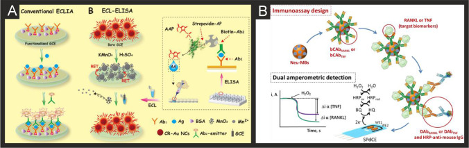
Determination of specific cardiac biomarkers (CBs) during the diagnosis and management of adverse cardiovascular events such as acute myocardial infarction (AMI) has become commonplace in emergency department (ED), cardiology and many other ward settings. Cardiac troponins (cTnT and cTnI) and natriuretic peptides (BNP and NT-pro-BNP) are the preferred biomarkers in clinical practice for the diagnostic workup of AMI, acute coronary syndrome (ACS) and other types of myocardial ischaemia and heart failure (HF), while the roles and possible clinical applications of several other potential biomarkers continue to be evaluated and are the subject of several comprehensive reviews. The requirement for rapid, repeated testing of a small number of CBs in ED and cardiology patients has led to the development of point-of-care (PoC) technology to circumvent the need for remote and lengthy testing procedures in the hospital pathology laboratories. Electroanalytical sensing platforms have the potential to meet these requirements. This review aims firstly to reflect on the potential benefits of rapid CB testing in critically ill patients, a very distinct cohort of patients with deranged baseline levels of CBs. We summarise their source and clinical relevance and are the first to report the required analytical ranges for such technology to be of value in this patient cohort. Secondly, we review the current electrochemical approaches, including its sub-variants such as photoelectrochemical and electrochemiluminescence, for the determination of important CBs highlighting the various strategies used, namely the use of micro- and nanomaterials, to maximise the sensitivities and selectivities of such approaches. Finally, we consider the challenges that must be overcome to allow for the commercialisation of this technology and transition into intensive care medicine.

Keywords: Biosensor; Cardiac biomarkers; Critically ill; Electroanalysis; Electrochemistry; Intensive care; Nanomaterial.


Conflict of interest statement

The authors declare no competing interests.

Figures

Fig. 1
Fig. 1
A) Schematic representation of sensing cTnI and cTnT biomarkers in a multiplexed sensor array format, utilising antibodies attached to ZnO nanorods. Reproduced with permission from ref [149]. Copyright Elsevier 2017. B) (i) Preparation process of the Ab2/Ag@SnO2 NFs signal probe; (ii) construction of the self-accelerated Ag@SnO2 NFs-based ECL immunosensor and (iii) proposed ECL mechanism for this system. Reproduced and adapted with permission from ref [151]. Copyright Elsevier 2018
Fig. 2
Fig. 2
A) Preparation procedure for the sandwich-type electrochemical cTnI immunosensor based on mesoporous Fe3O4. Reproduced with permission from [171]. Copyright Elsevier 2019. B) An illustration of (i) the specific target recognition and BFP release, and (ii) the ratiometric biosensing mechanism for cTnI using an MXENE based sensor. Reproduced and adapted with permission from [196]. Copyright Elsevier 2021
Fig. 3
Fig. 3
A) (i) Schematic diagram of the ECL biosensor array for the detection of three targets. (ii) Diagram and (iii) photograph of gold electrode array. B) ECL intensity-potential profiles for the determination of different concentration of myoglobin (i), cTnI (ii) and cTnT (iii). In (i): (ng/mL): (a) blank, (b) 0.050, (c) 0.10, (d) 0.25, (e) 0.50, (f) 0.75, (g) 1.0; In (ii) (pg/mL): (a) blank, (b) 1.0, (c) 2.0, (d) 4.0, (e) 6.0, (f) 8.0, (g) 10.0; In (iii) (ng/mL): (a) blank, (b) 0.50, (c) 0.75, (d) 1.0, (e) 2.0, (f) 3.0, (g) 4.0. Insert, calibration curve of Myo, cTnI and cTnT. Measurement conditions: 0.1 M PBS (pH 7.4) containing 50 mM TPA at a scan rate of 50 mV/s. Reproduced and adapted with permission from ref [163]. Copyright Elsevier 2018
Fig. 4
Fig. 4
A) Schematic diagram for the construction of the ratiometric immunosensor for detecting H-FABP based on Au/Pt nanocrystals and open-pored hollow carbon nanospheres. B) TEM images of the gold nanodendrites. C) Summary of the electrochemical sensing platform and DPV signal acquired at 0.001, 0.01, 0.1, 1.0, 10 and 200 ng mL−1 of H-FABP. Reproduced and adapted with permission from ref [205]. Copyright Elsevier 2021
Fig. 5
Fig. 5
A) Schematic illustration for the stepwise preparation of the biosensor for CK-MB based on creatine phosphate. Reproduced and adapted with permission from ref [210]. Copyright Elsevier 2014. B) Schematic for the fabrication of the label-free ECL CK-MB immunosensor based on an CNOS/Fe3O4/AuNPs/CS modified SPE. Reproduced and adapted with permission from ref [213]. Copyright Royal Society of Chemistry 2019. C) Schematic illustration of the AuPdCu NWNs-based electrochemical sensor for detecting CK-MB. Reproduced and adapted with permission from ref [214]. Copyright Elsevier 2021. D) Schematic overview of the porous PdPtCoNi@Pt-skin nanopolyhedra production and their incorporation into an electrochemical immunoassay for CK-MB. Reproduced and adapted with permission from ref [215]. Copyright Elsevier 2020
Fig. 6
Fig. 6
A) Schematic representation of the preparation and sensing of the CoPC@CNT based electrochemical immunoassay for BNP. Reproduced and adapted with permission from ref [238]. Copyright Wiley 2021. B) Schematic representation of the steps involved in the preparation of AuNPs-S-Phe-SPCEs (i) and HRP-anti-BNP-BNP-anti-BNP-AuNPs-S-Phe-SPCE immunosensor for the determination of BRP (ii). Reproduced and adapted with permission from ref [239]. Copyright Elsevier 2018
Fig. 7
Fig. 7
A) Schematic processes of the immunosensor fabrication based on Ab-HRP-AuNCs for the detection of NT-proBNP. Reproduced and adapted with permission from ref [241]. Copyright Elsevier 2011. B) Schematic showing the fabrication and detection methodology for the magnetoimmunosensor for the detection of NT-proBNP. Reproduced and adapted with permission from ref [243]. Copyright Elsevier 2013. C) Schematic description for the label-free NT-proBNP immunosensor based on ABEI/GNDs/chitosan/COOK-MWCNTs. Reproduced and adapted with permission from ref [247]. Copyright American Chemical Society 2015. D) Schematic of the production, composition and read-out of the ratiometric electrochemical immunoassay for NT-proBNP based on three dimensional PtCoNi hollow multi-branches/ferrocene-grafted-ionic liquid and Co–N-C nanosheets. Reproduced and adapted with permission from ref [261]. Copyright Elsevier 2021
Fig. 8
Fig. 8
A) Schematic of the sensor fabrication and representative DPV for the simultaneous immunosensing of multiple cytokines in serum. Reproduced and adapted with permission from ref [297]. Copyright American Chemical Society 2018. B) Schematic illustration of the immunosensor production and working mechanism based on porous carbon composites. Reproduced and adapted with permission from ref [311]. Copyright Elsevier 2021. C) Illustration of the synthesis procedure for NiCoO2@CeO2 NBs, the preparation of the electrocatalytic labels and the fabrication of the immunosensor. Reproduced and adapted with permission from reference [304]. Copyright American Chemical Society 2020. D) (i,ii) TEM images of NiCoO2@CeO2 NBs; (iii,iv) STEM image and elemental mapping of NiCoO2@CeO2 NBs. Reproduced and adapted with permission from ref [304]. Copyright American Chemical Society 2020
Fig. 9
Fig. 9
A) Schematic showing the fabrication of the gold wire sensor for CRP along with the detection strategy. Reproduced and adapted with permission from ref [323]. Copyright Elsevier 2019. B) Scanning electron microscopy images obtained at various magnifications of the Au/PC substrate used for the gold wire CRP sensor. Reproduced and adapted with permission from ref [323]. Copyright Elsevier 2019. C) Schematic of the preparation of rGO/Ni/PtNPs micromotors and their functionalisation with anti-CRP capture antibodies alongside SEM and EDX analysis (left), fluorescence microscopy images of the micromotors with and without streptavidin (right middle) and time-lapse images of the movement of the micromotors (right bottom). Reproduced and adapted with permission from ref [335]. Copyright American Chemical Society 2020
Fig. 10
Fig. 10
A) Schematic showing the bases of a conventional ECL-ELISA protocol and the ECL-ELISA protocol proposed by Peng and co-workers. Reproduced and adapted with permission from ref [356]. Copyright American Chemical Society 2019. B) Schematic showing the development of an MBs-based immune-platform for the dual amperometric detection of RANKL and TNF at dual SPCEs. Reproduced and adapted with permission from ref [358]. Copyright Elsevier 2020

Similar articles

Cited by

References

    1. McLean AS, Huang SJ, Nalos M, Tang B, Stewart DE. The confounding effects of age, gender, serum creatinine, and electrolyte concentrations on plasma B-type natriuretic peptide concentrations in critically ill patients. Crit Care Med. 2003;31:2611–2618. doi: 10.1097/01.CCM.0000094225.18237.20. - DOI - PubMed
    1. McLean AS, Tang B, Nalos M, Huang SJ, Stewart DE. Increased B-type natriuretic peptide (BNP) level is a strong predictor for cardiac dysfunction in intensive care unit patients. Anaesth Intensive Care. 2003;31:21–27. doi: 10.1177/0310057X0303100104. - DOI - PubMed
    1. Rudiger A, Singer M. Mechanisms of sepsis-induced cardiac dysfunction. Crit Care Med. 2007;35:1599–1608. doi: 10.1097/01.CCM.0000266683.64081.02. - DOI - PubMed
    1. Bak Z, Sjöberg F, Eriksson O, Steinvall I, Janerot-Sjoberg B. Cardiac dysfunction after burns. Burns. 2008;34:603–609. doi: 10.1016/j.burns.2007.11.013. - DOI - PubMed
    1. Venkata C, Kasal J. Cardiac Dysfunction in Adult Patients with Traumatic Brain Injury: A Prospective Cohort Study. Clin Med Res. 2018;16:57–65. doi: 10.3121/cmr.2018.1437. - DOI - PMC - PubMed