. 2022 Feb;24(2):55-61.
doi: 10.22074/cellj.2022.7622.

Differentiation of Human Wharton's Jelly Mesenchymal Stem Cells into SOX17 Expressing Cells Using a Wnt/ß-catenin Pathway Agonist on Polylactic Acid/Chitosan Nanocomposite Scaffold

Affiliations

Differentiation of Human Wharton's Jelly Mesenchymal Stem Cells into SOX17 Expressing Cells Using a Wnt/ß-catenin Pathway Agonist on Polylactic Acid/Chitosan Nanocomposite Scaffold

Elham Hoveizi et al. Cell J. 2022 Feb.

Abstract

Objective: The β-catenin signaling pathway promises the potential for differentiation of stem cells into definitive endoderm (DE) cells as precursors of beta cells. Therefore, it can be considered as an inducer for cell replacement therapies in diabetes. The main goal of this research is to successfully culture and induce differentiation of human Wharton's jelly mesenchymal stem cells (hWJMSCs) into Sox17-expressing cells using a Wnt/β-catenin pathway agonist (SKL2001) plus nanoparticles on a polylactic acid/chitosan (PLA/Cs) nanocomposite scaffold.

Materials and methods: In this experimental study, the nanocomposite was prepared through an electrospinning method and hWJMSCs were isolated through an explant technique. The morphology and the cell viability were evaluated by scanning electron microscopy (SEM) and 3-(4, 5- Dimethylthiazol-2)-2, 5-diphenyltetrazolium bromide (MTT) assay. Here, we present two differentiation protocols: the first one is induction with SKL2001; and the second one is with a combination of SKL2001 and zinc oxide nanoparticles (nZnO). Real-time quantitative reverse transcription (QRT-PCR) and immunocytochemistry analysis are carried out to examine the expression of specific markers in the differentiated cells.

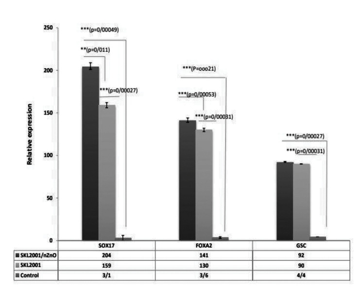
Results: The nanocomposite had appropriate biocompatibility for cell adhesion and growth. While the hWJMSCs cultured on the PLA/Cs scaffolds differentiated into DE cells in the presence of SKL2001, introducing nZnO to their environment increased the differentiation process. Analyses of DE-specific markers including SOX17, FOXA2, and gooscoid (GSC) genes in mRNA level, indicated significantly high levels of expression in the SKL2001/nZnO group, followed by SKL2001 group compared to the control.

Conclusion: Our results show the beneficial effects of the Wnt/β-catenin pathway agonist in three-dimensional (3D) cultures in cell replacement therapy for diabetes.

Keywords: Differentiation; Nanoparticles; Tissue Engineering; Wharton’s Jelly; Wnt/β-Catenin Pathway.


Conflict of interest statement

There is no conflict of interest in this study.

Figures

Fig.1
Fig.1
Characterization of human Wharton’s jelly mesenchymal stem cells (hWJMSCs). A. Typical observation of hWJMSCs under phase-contrast microscopy. B. Differentiation of hWJMSCs into osteoblasts as shown by Alizarin Red staining. C. Differentiation of hWJMSCs into adipocytes as shown by Oil Red O staining. D. Flow cytometric analysis of the isolated hWJMSCs for MSC (CD146, CD105, and CD90), and hematopoietic (CD34) and endothelial (CD31) markers (scale bar: 100 µm).
Fig.2
Fig.2
Scanning electron micrographs show the morphology of the plated human Wharton’s jelly mesenchymal stem cells (hWJMSCs) on polylactic acid/chitosan (PLA/Cs) scaffold on day 3 after seeding. A. The fibers of PLA/Cs scaffold were randomly entangled to form a strong, flexible, and porous 3D matrix. B. The fibers of PLA/Cs scaffold with higher magnification, C. The plated hWJMSCs on PLA/Cs scaffold, D. The plated hWJMSCs on PLA/Cs scaffold with higher magnification. Yellow arrows show the plated hWJMSCs on the scaffold.
Fig.3
Fig.3
MTT assay and morphological study of hWJMSCs by an inverted microscope. A. Formosan absorbance has been expressed as a measure of cell viability from the hWJMSCs cultured on nanocomposite scaffold for 6 days. B. Passage 1 hWJMSCs after 3 days in culture, C. Passage 3 hWJMSCs after 7 days, D. The cell viability assay using acridine orange/ ethidium bromide staining of hWJMSCs was performed on the cells cultured on PLA/Cs scaffold by fluorescent microscopy after 2 days (B, C, and D represent ×200, ×200, and ×100 magnification, respectively, scale bar: 100 µm). hWJMSCs; Human Wharton’s jelly mesenchymal stem cells, PLA/Cs; Polylactic acid/chitosan, and ***; P<0.05 and values are mean (n=3).
Fig.4
Fig.4
Quantitative expression analysis of DE cells derived from hWJMSCs cultured on PLA/Cs scaffold after 6 days. The results are collected from 3 independent experiments with 2 internal replicates per experiment. Differences observed were statistically significant when P≤0.05. Comparison of the gene expression levels of DE markers (FOXA2, SOX17, and GSC) in two experimental groups. hWJMSCs; Human Wharton’s jelly mesenchymal stem cells, PLA/Cs; Polylactic acid/chitosan, DE; Definitive endoderm, ***; P<0.001, and **; P<0.01 untreated cells were considered as a control group.
Fig.5
Fig.5
Immunocytochemistry performed for analyzing SOX17 and FOXA2 as endoderm-specific proteins by differentiated hWJMSCs on the scaffold after 6 days of culture. The staining of nuclei was performed by DAPI (×400 magnification, scale bar: 100 µm).

References

    1. Li XY, Wu SY, Leung PS. Human fetal bone marrow-derived mesenchymal stem cells promote the proliferation and differentiation of pancreatic progenitor cells and the engraftment function of islet-like cell clusters. Int J Mol Sci. 2019;20(17):4083–4083. - PMC - PubMed
    1. Cabezas J, Rojas D, Navarrete F, Ortiz R, Rivera G, Saravia F, et al. Equine mesenchymal stem cells derived from endometrial or adipose tissue share significant biological properties, but have distinctive pattern of surface markers and migration. Theriogenology. 2018;106:93–102. - PubMed
    1. Kamolz LP, Keck M, Kasper C. Wharton’s jelly mesenchymal stem cells promote wound healing and tissue regeneration. Stem Cell Res Ther. 2014;5(3):62–62. - PMC - PubMed
    1. Jurewicz E, Wyroba E, Filipek A. Tubulin-dependent secretion of S100A6 and cellular signaling pathways activated by S100A6-integrin β1 interaction. Cell Signal. 2018;42:21–29. - PubMed
    1. Ghorbani-Dalini S, Azarpira N, Sangtarash MH, Soleimanpour-Lichaei HR, Yaghobi R, Lorzadeh S, et al. Optimization of activin-A: a breakthrough in differentiation of human induced pluripotent stem cell into definitive endoderm. 3 Biotech. 2020;10(5):215–215. - PMC - PubMed

LinkOut - more resources