Review
. 2022 Mar:14:100231.
doi: 10.1016/j.mtbio.2022.100231. Epub 2022 Mar 5.

Multifunctional carbon nanomaterials for diagnostic applications in infectious diseases and tumors

Affiliations
Review

Multifunctional carbon nanomaterials for diagnostic applications in infectious diseases and tumors

Yang He et al. Mater Today Bio. 2022 Mar.

Abstract

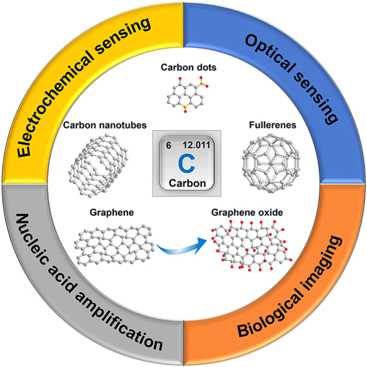
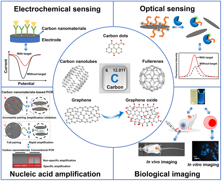
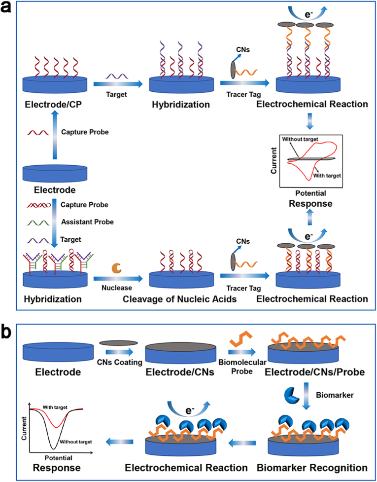
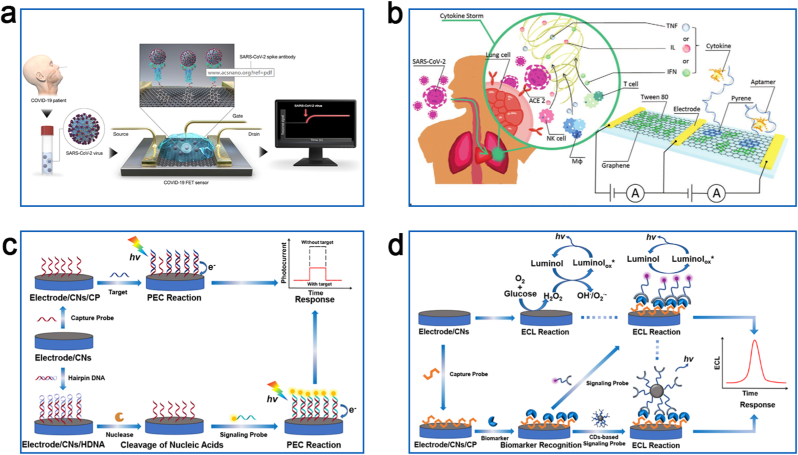
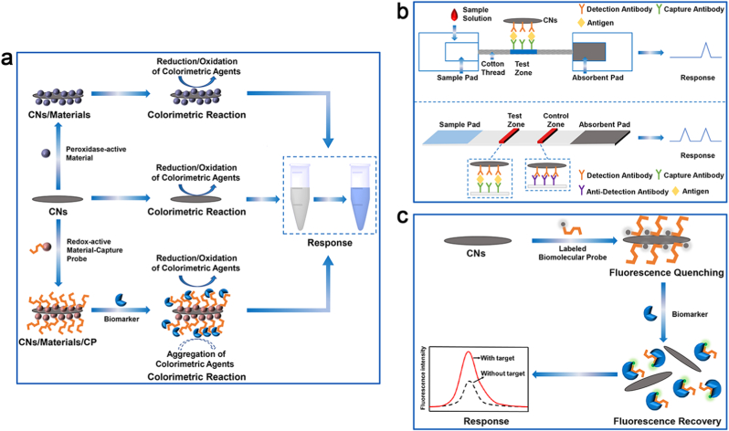
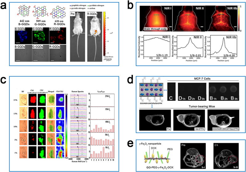
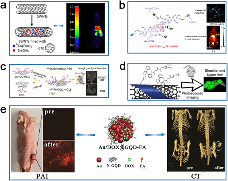
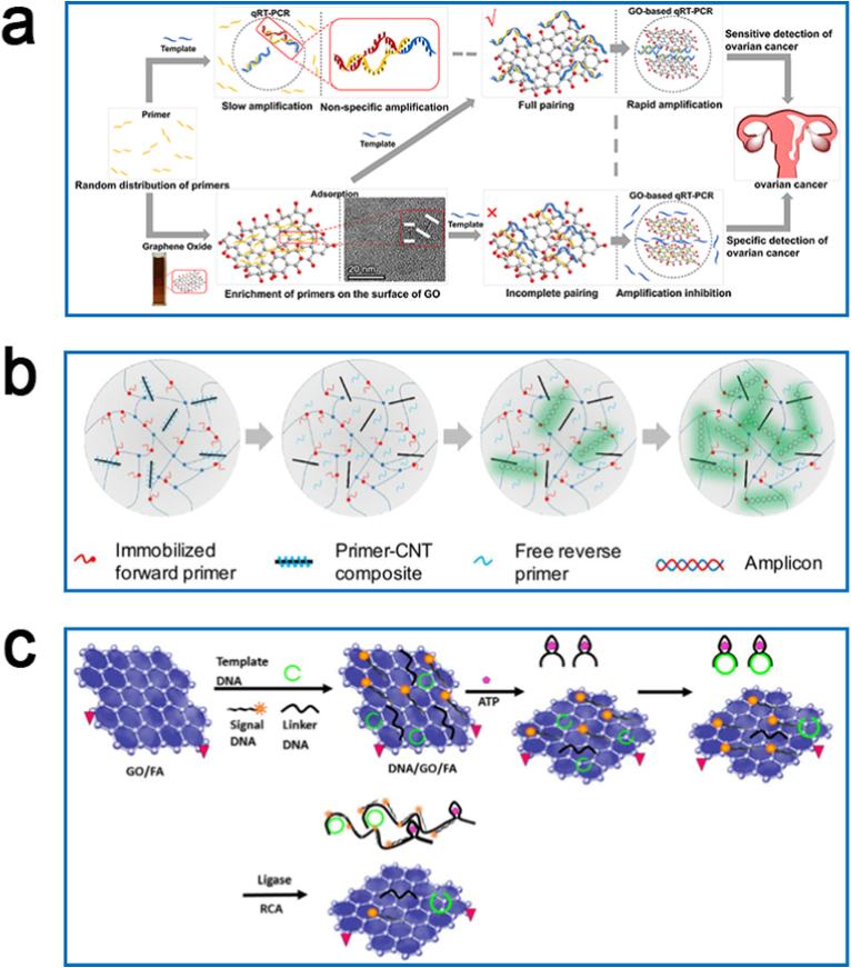
Infectious diseases (such as Corona Virus Disease 2019) and tumors pose a tremendous challenge to global public health. Early diagnosis of infectious diseases and tumors can lead to effective control and early intervention of the patient's condition. Over the past few decades, carbon nanomaterials (CNs) have attracted widespread attention in different scientific disciplines. In the field of biomedicine, carbon nanotubes, graphene, carbon quantum dots and fullerenes have the ability of improving the accuracy of the diagnosis by the improvement of the diagnostic approaches. Therefore, this review highlights their applications in the diagnosis of infectious diseases and tumors over the past five years. Recent advances in the field of biosensing, bioimaging, and nucleic acid amplification by such CNs are introduced and discussed, emphasizing the importance of their unique properties in infectious disease and tumor diagnosis and the challenges and opportunities that exist for future clinical applications. Although the application of CNs in the diagnosis of several diseases is still at a beginning stage, biosensors, bioimaging technologies and nucleic acid amplification technologies built on CNs represent a new generation of promising diagnostic tools that further support their potential application in infectious disease and tumor diagnosis.

Keywords: Carbon dots; Carbon nanotubes; Diagnosis; Diseases; Fullerenes; Graphene.


Conflict of interest statement

The authors declare that they have no known competing financial interests or personal relationships that could have appeared to influence the work reported in this paper.

Figures

Image 1
Graphical abstract
Fig. 1
Fig. 1
Carbon nanomaterials and their various applications in disease diagnosis. Electrochemical sensing. Optical sensing. Biological imaging, reproduced with permission from Refs. [14,131,143] (Copyrights 2020, Elsevier and Royal Society of Chemistry). Nucleic acid amplification, reproduced with permission from Ref. [162] (Copyright 2021, Elsevier).
Fig. 2
Fig. 2
Schematic presentation of conventional electrochemical biosensor for biomarker detection. (a) Schematic diagram of electrochemical biosensors with tracer tags constructed by CNs for biomarker detection. (b) Schematic presentation of electrochemical biosensors based on CN-modified electrodes for biomarker detection.
Fig. 3
Fig. 3
Schematic illustration of FET, PEC and ECL biosensors for biomarker detection. (a) Schematic diagram of the operating procedure of a graphene-based single-channel SARS-CoV-2 FET sensor. Reproduced with permission from Ref. [83] (Copyright 2020, American Chemical Society). (b) Schematic of the principle of a graphene-based dual-channel FET sensor for the detection of SARS-CoV-2-induced cytokine storm syndrome biomarkers. Reproduced with permission from Ref. [85] (Copyright 2021, Wiley). Sensing principles of PEC (c) and ECL (d) biosensors constructed based on CNs.
Fig. 4
Fig. 4
Schematic representation of optical biosensors for biomarker detection. Schematic diagrams of liquid-phase colorimetric biosensors (a), immunochromatographic biosensors (b) and fluorescent biosensors (c) for biomarker detection.
Fig. 5
Fig. 5
Optical imaging and MRI results of CNs discussed in the review. (a) Fluorescence images of HepG2 cells and tumor-bearing mice treated with GQDs of different emission wavelengths. Reproduced with permission from Ref. [131] (Copyright 2020, Elsevier). (b) NIR fluorescence images of the cerebrovasculature of mice treated with semiconductor SWCNTs. Reproduced with permission from Ref. [134] (Copyright 2015, Wiley). (c) Raman images of HepG2 cells incubated with SWCNT/Ag/AuNPs nanoprobe at different oxygen levels. Reproduced with permission from Ref. [138] (Copyright 2019, American Chemical Society) (d) MRI of MCF-7 ​cells and tumor-bearing mice treated with DOX/NGR-SWCNT/Gd-DTPA. Reproduced with permission from Ref. [141] (Copyright 2016, Taylor & Francis Group). Note: C, control group; D, Gd-DTPA; B, DOX/NGR-SWCNTs/Gd-DTPA. (e) MRI of H22 tumor-bearing nude mice treated with GO-PEG-γ-Fe2O3-DOX. The histogram demonstrates the temporal T2 signal of the tumor site acquired by region-of-interest (ROI) analysis. Reproduced with permission from Ref. [145] (Copyright 2018, Elsevier).
Fig. 6
Fig. 6
The non-optical imaging results of CNs discussed in the review. (a) PET image of healthy male Wistar rats treated with 64Cu@SWCNTs@β-D-glucan. Reproduced with permission from Ref. [147] (Copyright 2017, Elsevier). (b) Fluorescence image of U87MG cells treated with fluorescein-C60-PEG-cRGD and PET image of U87MG tumor-bearing mice treated with 64Cu-NOTA-C60-PEG-cRGD. Reproduced with permission from Ref. [148] (Copyright 2020, Elsevier). (c) SPECT/CT image and MRI of normal Balb/c mice treated with 99mTc/Gd-usNGO-PEG. Reproduced with permission from Ref. [149] (Copyright 2017, American Chemical Society). (d) PAI of bone localization in Balb/c mice treated with SWCNTs-BP-99mTc. Reproduced with permission from Ref. [153] (Copyright 2020, American Chemical Society). (e) PAI and CT image of tumor site in HeLa tumor-burdened nude mice injected via Au/DOX@GQD-FA. Reproduced with permission from Ref. [155] (Copyright 2019, Elsevier).
Fig. 7
Fig. 7
Schematic view of NAAT for biomarker detection. Schematic diagrams of GO-based qRT-PCR (a), CNT-based multiplex qRT-PCR (b) and GO-based RCA (c) for biomarker detection. Reproduced with permission from Refs. [162,166,168] (Copyright 2021 and 2017, Elsevier).

References

    1. Stadler K., Masignani V., Eickmann M., Becker S., Abrignani S., Klenk H.-D., Rappuoli R. SARS — beginning to understand a new virus. Nat. Rev. Microbiol. 2003;1:209–218. doi: 10.1038/nrmicro775. - DOI - PMC - PubMed
    1. Memish Z.A., Perlman S., van Kerkhove M.D., Zumla A. Middle East respiratory syndrome. Lancet. 2020;395:1063–1077. doi: 10.1016/S0140-6736(19)33221-0. - DOI - PMC - PubMed
    1. Zhu N., Zhang D., Wang W., Li X., Yang B., Song J., Zhao X., Huang B., Shi W., Lu R., Niu P., Zhan F., Ma X., Wang D., Xu W., Wu G., Gao G.F., Tan W. A novel coronavirus from patients with pneumonia in China, 2019. N. Engl. J. Med. 2020;382:727–733. doi: 10.1056/NEJMoa2001017. - DOI - PMC - PubMed
    1. Payandehpeyman J., Parvini N., Moradi K., Hashemian N. Detection of SARS-CoV-2 using antibody–antigen interactions with graphene-based nanomechanical resonator sensors. ACS Appl. Nano Mater. 2021;4:6189–6200. doi: 10.1021/acsanm.1c00983. - DOI - PubMed
    1. Mehra N.K., Jain A.K., Nahar M. Carbon nanomaterials in oncology: an expanding horizon. Drug Discov. Today. 2018;23:1016–1025. doi: 10.1016/j.drudis.2017.09.013. - DOI - PubMed

LinkOut - more resources