

An animal experiment study on the application of indocyanine green angiography in the harvest of multi-angiosome perforator flap
- PMID: 35280416
- PMCID: PMC8908162
- DOI: 10.21037/atm-22-220
An animal experiment study on the application of indocyanine green angiography in the harvest of multi-angiosome perforator flap
Abstract
Background: This study sought to explore the application value of indocyanine green angiography (ICGA) in the harvest of multi-angiosome perforator flap and the effect of low molecular weight heparin (LMWH) on the survival of postoperative flap.
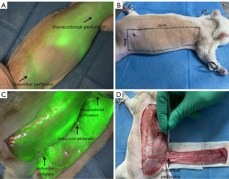
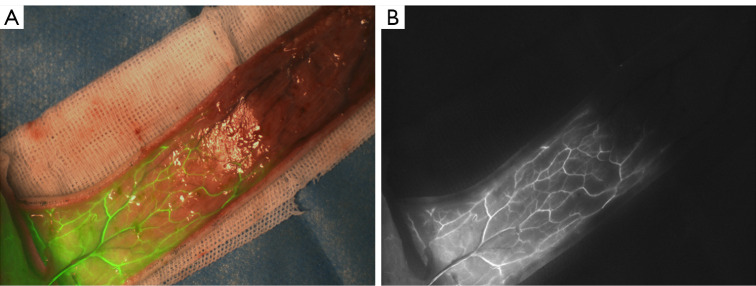
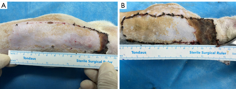
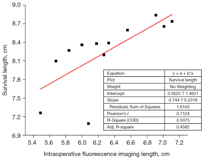
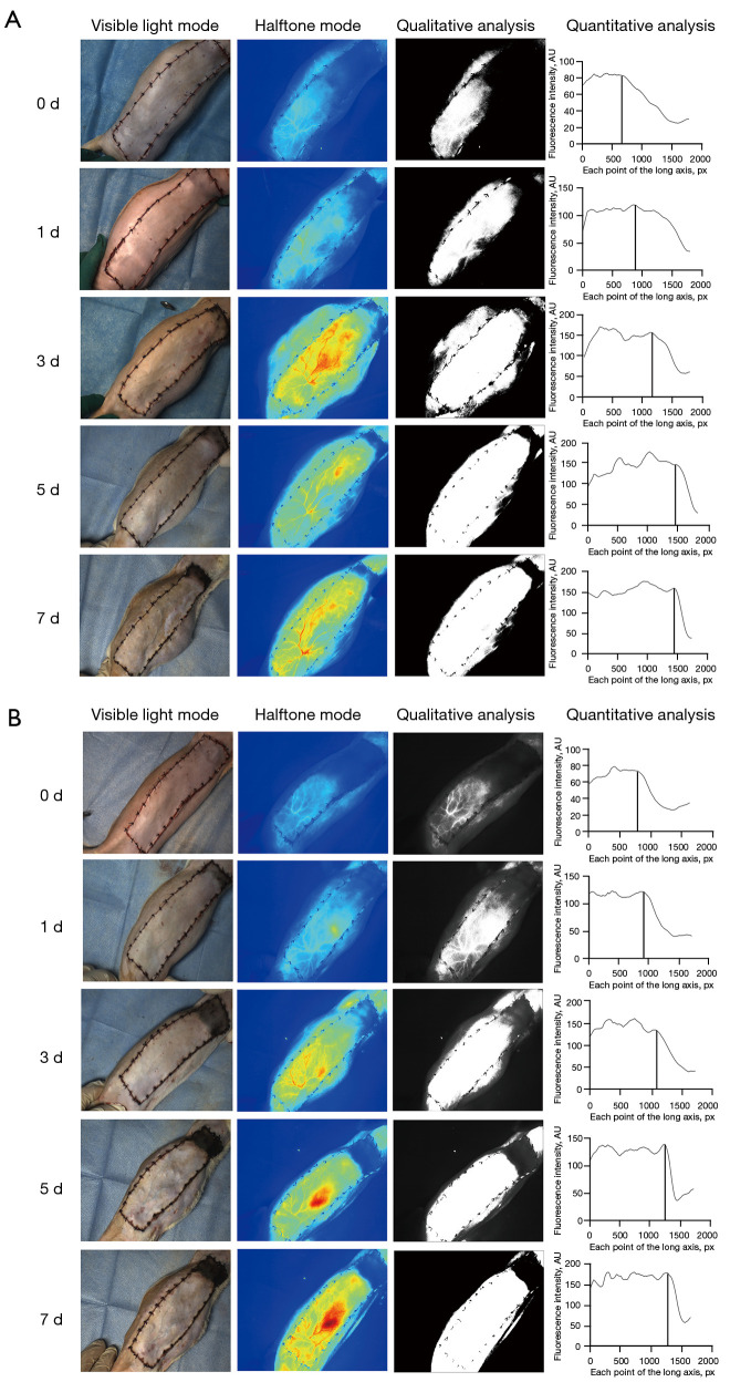
Methods: Twenty-four SD male rats were selected to construct a three-angiosome perforator flap model with the unilateral iliolumbar artery perforator. They were randomly divided into two groups: the control group was injected with indocyanine green (ICG) into the femoral vein during the operation, and the fluorescence signal was collected and quantitatively analyzed using Real-Time Image Guided System to determine the intraoperative fluorescence imaging length. The experimental group was injected subcutaneously with LMWH (400 U/kg) after 0.5 h postoperatively, and the control group was injected with the same amount of normal saline. The injection was repeated at the same time each day from 0 to 7 days postoperatively. After the flap was sutured in situ, ICGA was performed at 0, 1, 3, 5, and 7 days postoperatively to observe the vascular structure of the two groups of flaps. The flap survival length of the control group was counted at 7 days postoperatively, and the correlation between the intraoperative fluorescence imaging length and the survival length at 7 days postoperatively was calculated. The proportion of distal necrosis of the flaps between the two groups was compared at 7 days postoperatively.
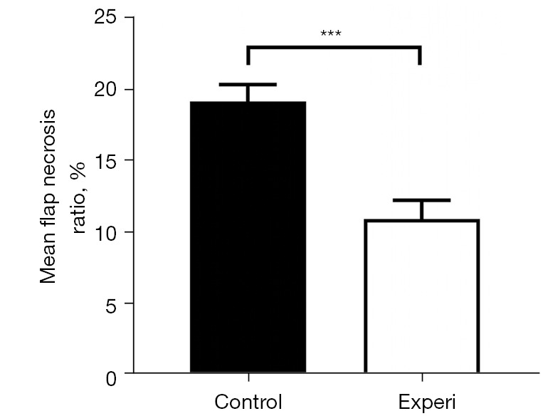
Results: The average length of intraoperative fluorescence imaging in the control group was 6.29±0.50 cm, and the survival length of the flap at 7 days postoperatively was 8.24±0.52 cm. The actual survival length was higher than the intraoperative fluorescence imaging length, with a ratio of 1.31±0.08. The difference was statistically significant (P<0.05). At 7 days postoperatively, the flap necrosis ratio of experimental group and control group were 10.92%±1.30% and 19.11%±1.19%, and the flap necrosis ratio of experimental group was lower than that of control group (P<0.001).
Conclusions: ICGA can locate the position of perforator, and can be used to predict and observe the length of distal survival of multi-angiosome perforator flap postoperatively. LMWH can promote the distal survival of flap and reduce flap necrosis.
Keywords: Indocyanine green angiography; angiosome; limit length; low molecular weight heparin; perforator flap.
2022 Annals of Translational Medicine. All rights reserved.
Conflict of interest statement
Conflicts of Interest: All authors have completed the ICMJE uniform disclosure form (available at https://atm.amegroups.com/article/view/10.21037/atm-22-220/coif). The authors have no conflicts of interest to declare.
Figures
Similar articles
-
[Intraoperative verification of a perforator flap vascularization by indocyanine green angiography].Ann Chir Plast Esthet. 2014 Feb;59(1):70-5. doi: 10.1016/j.anplas.2013.04.002. Epub 2013 Jul 26. Ann Chir Plast Esthet. 2014. PMID: 23896575 French.
-
Flap warming improves intraoperative indocyanine green angiography (ICGA) assessment of perfusion. An experimental study.J Plast Reconstr Aesthet Surg. 2019 Jul;72(7):1150-1156. doi: 10.1016/j.bjps.2019.03.014. Epub 2019 Mar 28. J Plast Reconstr Aesthet Surg. 2019. PMID: 30952589
-
[Effects of pretreatment with dimethyloxalylglycine on the survival of multi-territory perforator flap in rat and related mechanism].Zhonghua Shao Shang Za Zhi. 2016 Jul 20;32(7):396-401. doi: 10.3760/cma.j.issn.1009-2587.2016.07.003. Zhonghua Shao Shang Za Zhi. 2016. PMID: 27464629 Chinese.
-
Dynamic perfusion assessment during perforator flap surgery: an up-to-date.Clujul Med. 2015;88(3):293-7. doi: 10.15386/cjmed-484. Epub 2015 Jul 1. Clujul Med. 2015. PMID: 26609259 Free PMC article. Review.
-
Optimizing Indocyanine Green Fluorescence Angiography in Reconstructive Flap Surgery: A Systematic Review and Ex Vivo Experiments.Surg Innov. 2020 Feb;27(1):103-119. doi: 10.1177/1553350619862097. Epub 2019 Jul 26. Surg Innov. 2020. PMID: 31347468
References
LinkOut - more resources
Full Text Sources