. 2022 Feb;14(2):238-246.
doi: 10.21037/jtd-21-1242.

Development of a minimally invasive pulmonary porcine embolism model via endobronchial ultrasound

Affiliations

Development of a minimally invasive pulmonary porcine embolism model via endobronchial ultrasound

Terunaga Inage et al. J Thorac Dis. 2022 Feb.

Abstract

Background: Current massive pulmonary embolism (PE) animal models use central venous access to deliver blood clots, which have features of random clot distribution and potentially fatal hemodynamic compromise. A clinically relevant preclinical model for generating pulmonary emboli in a more controlled fashion would be of value for a variety of research studies, including initial evaluation of novel therapeutic approaches. Endobronchial ultrasound-guided transbronchial needle injection (EBUS-TBNI) is a newly established approach for peri-tracheal/bronchial targets. The purpose of the present work was to establish a minimally invasive PE model in swine via a transbronchial approach.

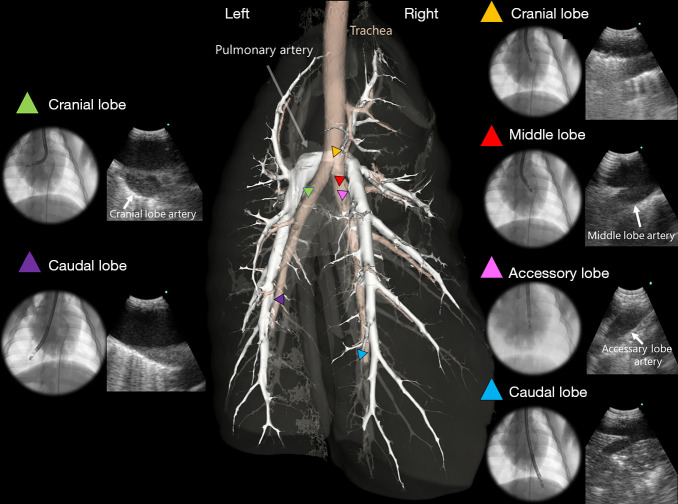
Methods: In anesthetized Yorkshire pigs, a 21-G EBUS-guided transbronchial needle aspiration (EBUS-TBNA) needle was introduced into the pulmonary artery under EBUS guidance. Autologous blood clots were administered into the right and left lower pulmonary arteries sequentially (PE1 and PE2, respectively). Hemodynamic and biochemical responses were evaluated.

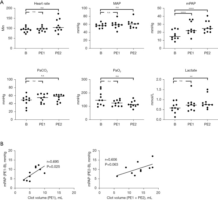
Results: Ten pigs were evaluated; all 20 blood clots (6.3±1.9 mL) were successfully injected. After injection, mean pulmonary artery pressure (mPAP; mmHg) increased (baseline: 16.6±5.6 vs. PE1: 24.5±7.6, P<0.0001 vs. PE2: 26.9±6.7, P<0.0001), and a positive correlation was observed between clot volume and change in mPAP (PE1: r=0.69, P=0.025; PE1 + PE2: r=0.60, P=0.063). Mean arterial pressure (MAP; mmHg) (baseline: 57.5±5.1 vs. PE1: 59.0±9.1, P=0.918 vs. PE2: 60.9±9.6, P=0.664) remained stable. No complications were observed.

Conclusions: EBUS allows minimally invasive, precise, and reliable generation of pulmonary emboli in pigs. This model may serve as an important tool for new PE-related diagnostic and therapeutic research.

Keywords: Pulmonary embolism (PE); endobronchial ultrasound (EBUS); swine.


Conflict of interest statement

Conflicts of Interest: All authors have completed the ICMJE uniform disclosure form (available at https://jtd.amegroups.com/article/view/10.21037/jtd-21-1242/coif). KY reports that this work was supported by a research grant from Olympus Corporation, and KY is a consultant for Olympus. The other authors have no conflicts of interest to declare.

Figures

Figure 1
Figure 1
Ex vivo autologous blood clot formation. Blood (A) is incubated 2 h at 37 °C in a syringe to encourage the formation of solid clot (B). Clot ‘aliquots’ are then transferred into 1 mL syringes (C) for administration via a 21-G EBUS-TBNA needle (D). EBUS-TBNA, endobronchial ultrasound-guided transbronchial needle aspiration.
Figure 2
Figure 2
Study protocol.
Figure 3
Figure 3
EBUS-TBNI for generation of pulmonary emboli. (A) EBUS B-mode and (B) Doppler images identify the pulmonary artery. (C) Puncture of the artery by the 21-G EBUS-TBNA needle is monitored using B-mode. (D) Intravascular clot injection is monitored in real-time using EBUS; expelled clot is noted by the white arrow. (E) After injection, a large clot is seen on EBUS B-mode (yellow box and arrow). (F) Disrupted arterial flow is confirmed by EBUS Doppler (red arrow marks proximal edge of embolus). EBUS-TBNI, endobronchial ultrasound-guided transbronchial needle injection; EBUS-TBNA, endobronchial ultrasound-guided transbronchial needle aspiration.
Figure 4
Figure 4
Radiological and pathological evaluation of pulmonary emboli. (A) Pulmonary artery angiography demonstrates a filling defect (red arrow), confirming clot formation. (B) Contrast-enhanced cone beam computed tomography similarly demonstrates a filling defect (red arrow; pulmonary artery catheter indicated by blue arrow). (C) Embolization of the pulmonary artery was confirmed by postmortem examination; this specific embolus followed injection of 7 mL clot into the right lower lobe pulmonary artery. (D) Pathological evaluation reveals large arterial emboli (red arrows) with adjacent regions of hemorrhagic pulmonary infarction (yellow arrows); staining method: hematoxylin and eosin staining; scale bar corresponds to 4 mm (left) and 300 µm (right).
Figure 5
Figure 5
Changes in hemodynamics and biochemistry post-clot injection. (A) HR, MAP, mPAP, PaCO2, PaO2, and lactate are shown from B, PE1 and PE2. Data presented as mean ± SEM. (B) Injected clot volume (PE1 or PE1 + PE2) correlates with change in mPAP. **, P<0.01; ****, P<0.0001. B, baseline; PE1, post-clot injection 1; PE2, post-clot injection 2. n.s., not significant; MAP, mean arterial pressure; mPAP, mean pulmonary artery pressure; PaCO2, partial pressure of arterial carbon dioxide; PaO2, partial pressure of arterial oxygen; HR, heart rate; SEM, standard error of the mean.
Figure 6
Figure 6
3D reconstruction of pig lung anatomy. Virtual model of the bronchial and arterial tree demonstrate pulmonary artery access to the right cranial lobe artery (yellow), middle lobe artery (red), accessary lobe artery (pink), right caudal lobe artery (blue), left cranial lobe artery (green) and left caudal lobe artery (purple) using EBUS. EBUS, endobronchial ultrasound.

References

    1. Barbash IM, Schenke WH, Halabi M, et al. Experimental model of large pulmonary embolism employing controlled release of subacute caval thrombus in swine. J Vasc Interv Radiol 2011;22:1471-7. 10.1016/j.jvir.2011.06.011 - DOI - PMC - PubMed
    1. Schultz J, Andersen A, Gade IL, et al. A porcine in-vivo model of acute pulmonary embolism. Pulm Circ 2018;8:2045893217738217. 10.1177/2045893217738217 - DOI - PMC - PubMed
    1. Kjaergaard B, Kristensen SR, Risom M, et al. A porcine model of massive, totally occlusive, pulmonary embolism. Thromb Res 2009;124:226-9. 10.1016/j.thromres.2009.01.010 - DOI - PubMed
    1. Zhao LB, Jia ZY, Lu GD, et al. Establishment of a canine model of acute pulmonary embolism with definite right ventricular dysfunction through introduced autologous blood clots. Thromb Res 2015;135:727-32. 10.1016/j.thromres.2015.01.016 - DOI - PubMed
    1. Khan F, Anker CJ, Garrison G, et al. Endobronchial ultrasound-guided transbronchial needle injection for local control of recurrent non-small cell lung cancer. Ann Am Thorac Soc 2015;12:101-4. 10.1513/AnnalsATS.201408-358BC - DOI - PubMed