. 2022 Jan 24;18(4):1521-1538.
doi: 10.7150/ijbs.66477. eCollection 2022.

Caspase-1-mediated extracellular vesicles derived from pyroptotic alveolar macrophages promote inflammation in acute lung injury

Affiliations

Caspase-1-mediated extracellular vesicles derived from pyroptotic alveolar macrophages promote inflammation in acute lung injury

Xichun Qin et al. Int J Biol Sci. .

Abstract

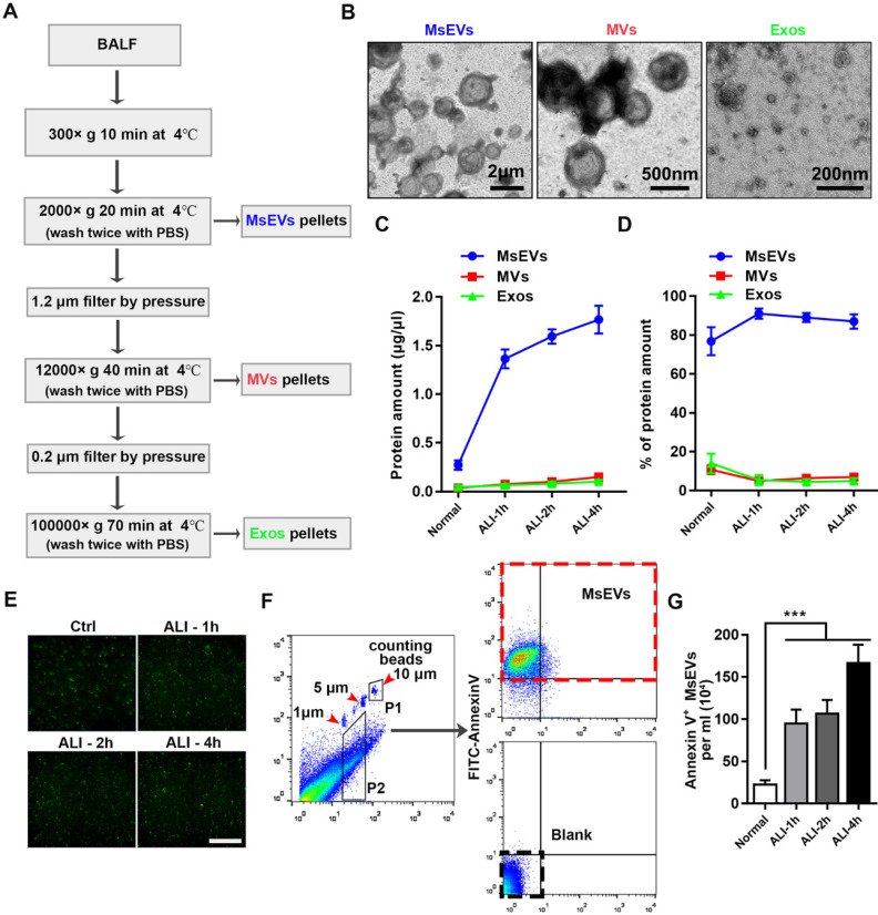
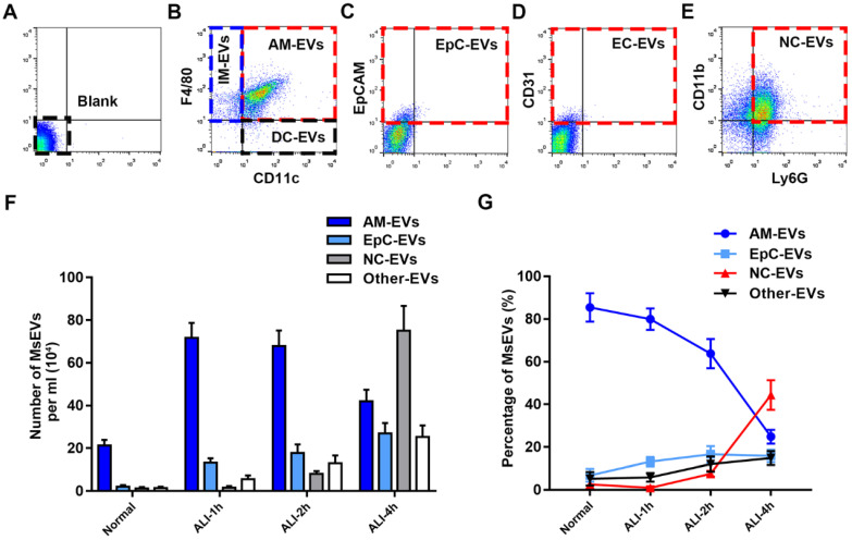
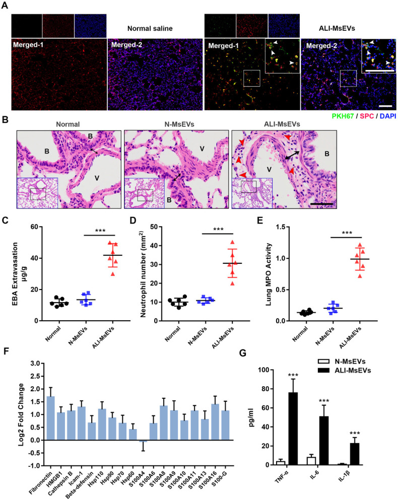
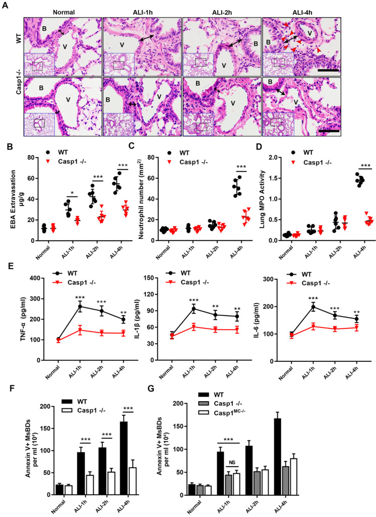
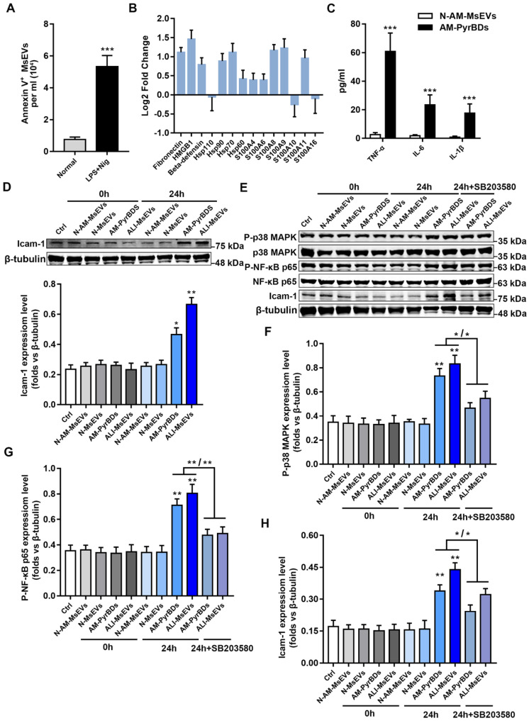
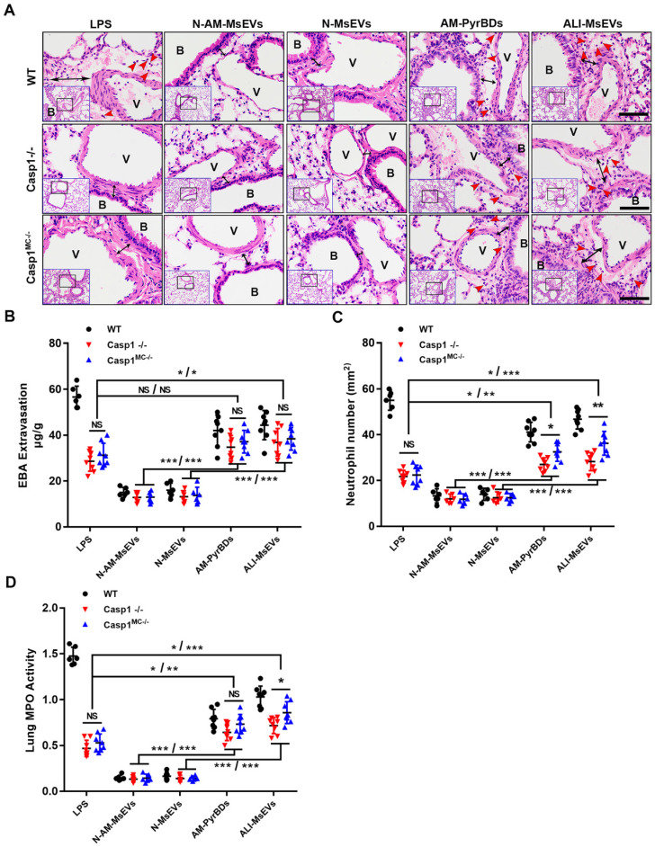
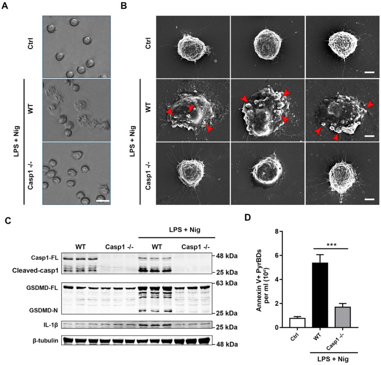
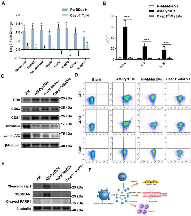
The occurrence and development of acute lung injury (ALI) involve a variety of pathological factors and complex mechanisms. How pulmonary cells communicate with each other and subsequently trigger an inflammatory cascade remains elusive. Extracellular vesicles (EVs) are a critical class of membrane-bound structures that have been widely investigated for their roles in pathophysiological processes, especially in immune responses and tumor progression. Most of the current knowledge of the functions of EVs is related to functions derived from viable cells (e.g., microvesicles and exosomes) or apoptotic cells (e.g., apoptotic bodies); however, there is limited understanding of the rapidly progressing inflammatory response in ALI. Herein, a comprehensive analysis of micron-sized EVs revealed a mass production of 1-5 μm pyroptotic bodies (PyrBDs) release in the early phase of ALI induced by lipopolysaccharide (LPS). Alveolar macrophages were the main source of PyrBDs in the early phase of ALI, and the formation and release of PyrBDs were dependent on caspase-1. Furthermore, PyrBDs promoted the activation of epithelial cells, induced vascular leakage and recruited neutrophils through delivery of damage-associated molecular patterns (DAMPs). Collectively, these findings suggest that PyrBDs are mainly released by macrophages in a caspase-1-dependent manner and serve as mediators of LPS-induced ALI.

Keywords: acute lung injury; alveolar macrophage; caspase-1; extracellular vesicles; pyroptosis.


Conflict of interest statement

Competing Interests: The authors have declared that no competing interest exists.

Figures

Figure 1
Figure 1
Mass release of micrometer-sized extracellular vesicles in the early phase of ALI C57BL/6J mice induced by LPS. (A) Protocol to harvest micron-sized vesicles, microvesicles, and exosomes. BALF was harvested from mice instilled or not instilled with LPS, and extracellular vesicles were separated by serial centrifugation. The pellets were washed with PBS and then centrifuged twice to remove soluble factors or impurities outside the vesicles. (B) Representative transmission electron micrograph of extracellular vesicles pellets derived from differential centrifugation. Bar (from left to right) = 2 μm, 500 nm, 200 nm. (C) Protein concentration of three types of EVs and (D) the percentages of each type of EVs. (E) Immunostaining showing positive expression of FITC-Annexin V in the pellets. The pellets were incubated with FITC-labeled Annexin V+ for 15 min, washed twice with PBS, and observed under a fluorescence microscope. (F) The number of MsEVs was determined by flow cytometry. One- and 5-μm-diameter beads and 10-μm counting beads were used to gate 1-5 μm-sized EVs. Annexin V+ vesicles were counted as MsEVs (P2 * Annexin V+ rate / P1 * number of counting beads). Data are expressed as mean ± SD, n = 6. ***P < 0.001.
Figure 2
Figure 2
Characterization of MsEVs from different cell types. (A-E) The origin of BALF MsEVs classified by flow cytometry. CD11c+ and F4/80+ events designate alveolar macrophages; EpCAM+—epithelial cells; other events (such as CD31+ for endothelial cells, CD11c+ F4/80- for dendritic cells, CD11c- F4/80+ for interstitial macrophages) were pooled into the Other group. (F and G) Quantitative measurement of MsEVs. MsEVs from different cell sources were detected at different time points in the early phase of the ALI model (n=6). Data are expressed as the mean ± SD. AM, alveolar macrophage; IM, interstitial macrophage; DC, dendritic cell; EpC, epithelial cell; NC, neutrophil; EC, endothelial cell.
Figure 3
Figure 3
MsEVs derived from alveolar macrophages in the early stage of ALI encapsulate DAMPs and mediate inflammation. (A) After tracheal drip of PKH67-labeled PyrBDs for 4 h, immunofluorescent staining showed that PKH67 co-localized with SPC, indicating the uptake of PyrBDs by lung epithelial cells. The white arrow points to undegraded PyrBDs (green); bar = 100 μm. The number of EVs is one million per mouse. SPC (red); DAPI (blue). (B) H&E-stained cross-section of the lung from N-MsEVs (MsEVs from the normal group) and ALI-MsEVs (MsEVs from the ALI-1h group) exposed mice at 4 h shows interstitial edema (indicated by black arrows). The red arrow points to neutrophils. Scale bars: 50 μm. B, trachea; V, blood vessel. Images are representative of six animals. (C) Assessment of the degree of lung vascular leakage (EBA extravasation). (D) Quantitative analysis of neutrophil infiltration in the lungs. (E) Quantitative analysis of lung tissue MPO activity. (F) PRM for whole-protein analysis of MsEVs, from which DAMP-related molecular proteins were screened. EVs pellets were isolated from BALF collected from control mouse or 1 h post-LPS. The results are expressed as log2 fold change (n = 5). (G) TNF-α, IL-6, and IL-1β content of MsEVs. The EVs pellets were ultrasonically broken, the supernatant was obtained by centrifugation at 14,000g, and cytokine content was detected by ELISA kit. Compared with N-MsEVs, ALI-MsEVs contain high levels of TNF-α, IL-6, and IL-1β. Data are expressed as the mean ± SD, n = 6. ***P < 0.001.
Figure 4
Figure 4
Requirement for caspase-1 in mice in mediating inflammatory damage and release of MsEVs. (A) H&E-stained cross-section of the lung from LPS-exposed WT and Casp1-/- mice at 1, 2, and 4 h shows interstitial edema (indicated by black arrows) in the ALI groups. The red arrow points to neutrophils. Scale bars: 50 μm. B, trachea; V, blood vessel. Images are representative of six animals. Quantitative analysis of (B) lung vascular leakage (EBA extravasation), (C) neutrophil infiltration and (D) lung tissue MPO activity. Lack of caspase-1 significantly reduced LPS-induced vascular leakage and neutrophil infiltration. (E) Quantification of cytokines. TNF-α, IL-1β, and IL-6 expression increased in the WT groups compared with the Casp1-/- groups (n=6). Data are expressed as the mean ± SD. **P < 0.01; ***P < 0.001. (F) The number of MsEVs was determined by flow cytometry. The same dose of LPS was instilled into the trachea; compared with the WT ALI model group, the amount of MsEVs significantly decreased in Casp1-/- mice. (G) Specifically knocking out macrophage caspase-1 in mice, the release of MsEVs triggered by LPS was significantly reduced. Data are expressed as the mean ± SD. NS, no significant difference; **P < 0.01; ***P < 0.001.
Figure 5
Figure 5
PyrBDs activate the p38 MAPK pathway to promote MLE-12 cell activation. (A) The number of MsEVs was determined by flow cytometry. With the occurrence of pyroptosis, alveolar macrophages derived a large number of MsEVs (PyrBDs). Data are expressed as the mean ± SD, n = 3. (B) PRM for whole-protein analysis of PyrBDs, from which DAMP-related molecular proteins were screened. The results are expressed as log2 fold change (n = 4). (C) TNF-α, IL-6, and IL-1β content of PyrBDs. Compared with MsEVs derived from normal alveolar macrophages (N-AM-MsEVs), MsEVs derived from pyroptotic alveolar macrophages (AM-PyrBDs) contained high levels of TNF-α, IL-6, and IL-1β. Data are expressed as the mean ± SD, n = 6, ***P < 0.001. (D) Immunoblot analysis for icam-1. AM-PyrBDs and ALI-MsEVs (derived from ALI-1h mice) groups showed the promoted expression of icam-1 in MLE-12 cells. N-MsEVs, isolated from BALF of normal mouse; N-AM-MsEVs, derived from normal alveolar macrophages; (E) Immunoblot analysis of phospho-p38 MAPK, p38 MAPK, phospho-NF-κB p65, NF-κB p65, and icam-1. Quantification of the related protein expression showed significant upregulation of phospho-p38 MAPK (F), phospho-NF-κB p65 (G), and icam-1 (H), which was reversed by the inhibitor SB 203580. Data are expressed as the mean ± SD, n = 3. *P < 0.05; **P < 0.01.
Figure 6
Figure 6
PyrBDs induce vascular leakage and recruit neutrophils. (A) H&E-stained cross-section of the lung from LPS-exposed or PyrBDs treated WT, Casp1-/-, and Casp1MC-/- mice at 4 h showed that PyrBDs were able to cause extensive neutrophils infiltration of the lung interstitium. The red arrow points to neutrophils; the black arrow indicates interstitial edema; Bar = 50 μm. B, trachea; V, blood vessel. Quantitative analysis of (B) lung vascular leakage (EBA extravasation), (C) neutrophil infiltration, and (D) lung tissue MPO activity. Compared with the N-MsEV and N-AM-MsEV groups, the PyrBD groups showed more severe vascular leakage and neutrophil infiltration. In addition, PyrBDs reversed the resistance of caspase-1 deficiency to LPS to a certain extent. N-AM-MsEVs, normal alveolar macrophages MsEVs; N-MsEVs, normal mouse MsEVs; AM-PyrBDs, pyroptotic alveolar macrophages MsEVs; ALI-MsEVs, ALI-1h mice MsEVs. Data are expressed as the mean ± SD, NS, no significant difference; *P < 0.05; **P < 0.01; ***P < 0.001.
Figure 7
Figure 7
Caspase-1 mediates the formation and release of PyrBDs in macrophages. (A) Observation of the pyroptosis of alveolar macrophages by microscope. (B) Representative scanning electronic microscopy (SEM) images of alveolar macrophages treated with LPS and Nig. Alveolar macrophages from the WT mice showed swelling, release of cytoplasmic content, plasma membrane bubbles, and adhesion of micron-sized vesicles. In the Casp1-/- group, no similar morphological changes were observed. Bar = 5 μm. (C) Immunoblot analysis for pyroptosis-related proteins, caspase-1 full length (Casp1-FL), cleaved caspase-1, GSDMD full length (GSDMD-FL), GSDMD-N-terminal (GSDMD-N), and IL-1β. Data are expressed as the mean ± SD. (D) Lack of caspase-1 led to a decrease in PyrBDs released by alveolar macrophages. Data are expressed as the mean ± SD, ***P < 0.001.
Figure 8
Figure 8
PyrBDs distinctly express cleaved caspase-1 and its substrate proteins. (A) PRM for whole protein analysis of PyrBDs, from which DAMP-related molecular proteins were screened. Loss of caspase-1 led to a decrease in DAMP content. The results are expressed as log2 fold change (n = 4), *P < 0.05. (B) TNF-α, IL-6, and IL-1β content of PyrBDs. Loss of caspase-1 led to decreased expression of inflammatory factors. Data are expressed as the mean ± SD, n = 6, ***P < 0.001. (C) Immunoblot analysis for CD9, CD63, CD81, Histone 3, and Lamin A/C in PyrBDs. (D) Flow cytometry detected the expression of transmembrane proteins CD9, CD63, and CD81 in PyrBDs. (E) Immunoblot analysis for cleaved caspase-1, GSDMD-N, and cleaved PARP1 in PyrBDs. (F) Schematic diagram demonstrating that PyrBDs derived from pyroptotic alveolar macrophages specifically express cleaved caspase-1 and carry DAMPs, mediate epithelial cells activation, increase vascular permeability, and recruit neutrophils.

References

    1. Bhattacharya J, Matthay MA. Regulation and repair of the alveolar-capillary barrier in acute lung injury. Annu Rev Physiol. 2013;75:593–615. - PubMed
    1. McVey MJ, Steinberg BE, Goldenberg NM. Inflammasome Activation in Acute Lung Injury. Am J Physiol Lung Cell Mol Physiol. 2020. - PubMed
    1. Force ADT, Ranieri VM, Rubenfeld GD, Thompson BT, Ferguson ND, Caldwell E. et al. Acute respiratory distress syndrome: the Berlin Definition. JAMA. 2012;307:2526–33. - PubMed
    1. Bellani G, Laffey JG, Pham T, Fan E, Brochard L, Esteban A. et al. Epidemiology, Patterns of Care, and Mortality for Patients With Acute Respiratory Distress Syndrome in Intensive Care Units in 50 Countries. JAMA. 2016;315:788–800. - PubMed
    1. Poon IKH, Parkes MAF, Jiang L, Atkin-Smith GK, Tixeira R, Gregory CD. et al. Moving beyond size and phosphatidylserine exposure: evidence for a diversity of apoptotic cell-derived extracellular vesicles in vitro. J Extracell Vesicles. 2019;8:1608786. - PMC - PubMed

Publication types