. 2022 Feb 25:9:741989.
doi: 10.3389/fmed.2022.741989. eCollection 2022.

Altered Differentiation and Inflammation Profiles Contribute to Enhanced Innate Responses in Severe COPD Epithelium to Rhinovirus Infection

Affiliations

Altered Differentiation and Inflammation Profiles Contribute to Enhanced Innate Responses in Severe COPD Epithelium to Rhinovirus Infection

Hong Guo-Parke et al. Front Med (Lausanne). .

Abstract

Background: Respiratory viral infections are closely associated with COPD exacerbations, hospitalisations, and significant morbidity and mortality. The consequences of the persisting inflammation and differentiation status in virus associated severe disease is not fully understood. The aim of this study was to evaluate barrier function, cellular architecture, the inflammatory response in severe COPD bronchial epithelium to human rhinovirus (HRV) induced pathological changes and innate immune responses.

Methods: Well-differentiated primary bronchial epithelial cells (WD-PBECs) derived from severe COPD patients and age-matched healthy controls were cultured in the air-liquid interface (ALI) model. The differentiation phenotype, epithelial barrier integrity, pathological response and cytokine secreting profile of these cultures before and after HRV infection were investigated.

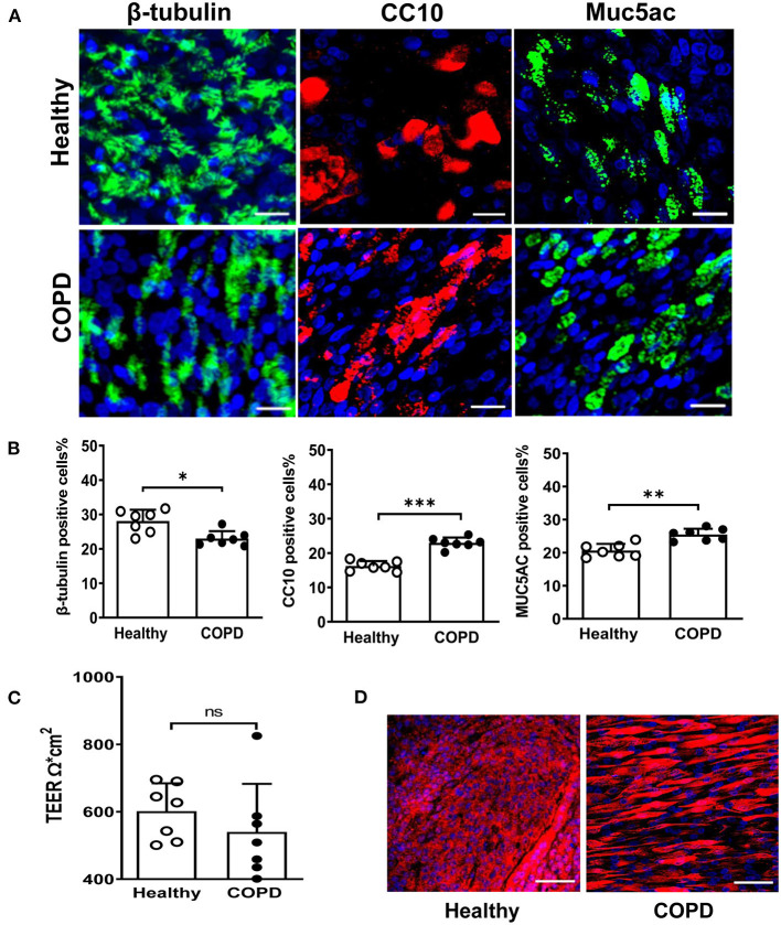
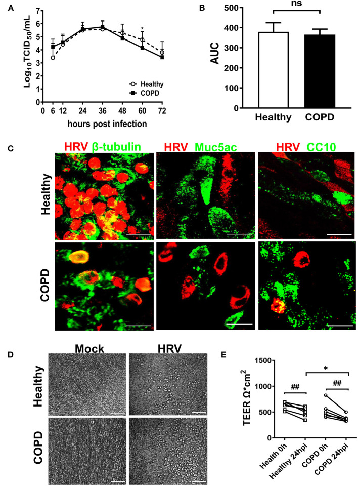
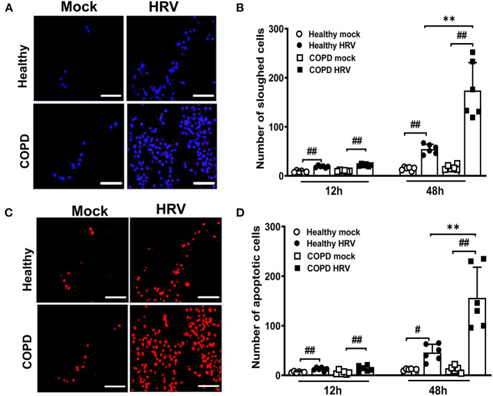
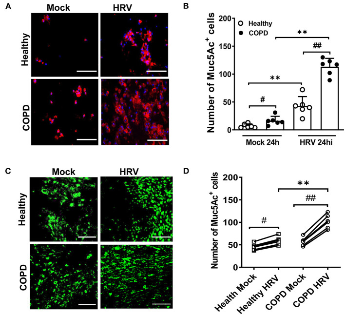
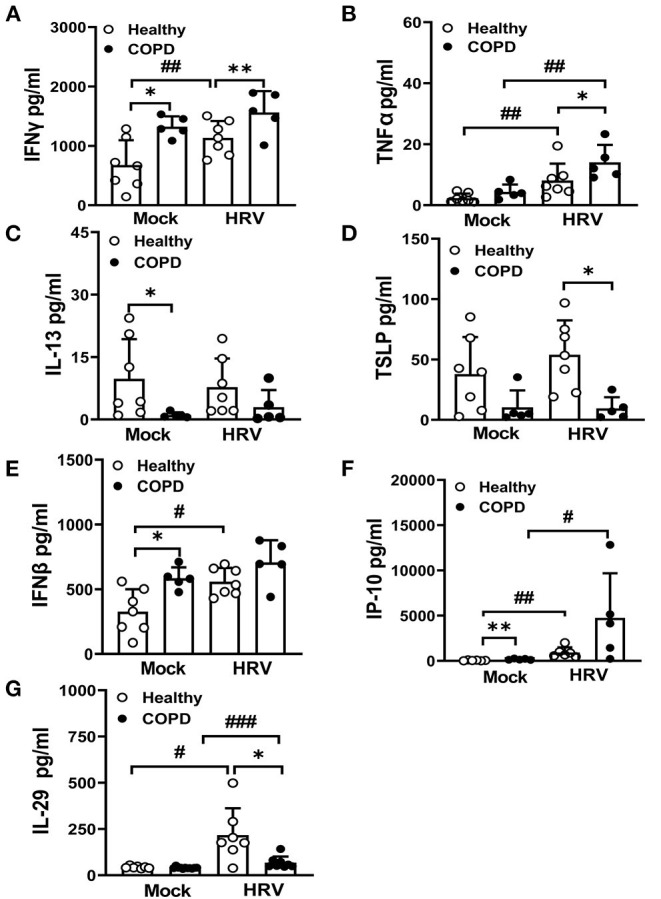
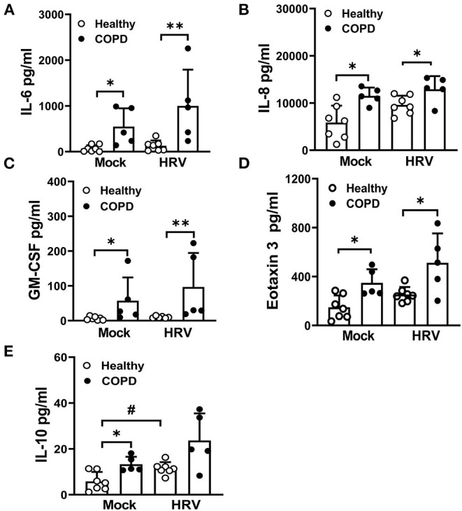
Results: WD-PBECs derived from severe COPD patients showed aberrant epithelium differentiation with a decreased proportion of ciliated cells but increased numbers of club cells and goblet cells compared with healthy controls. Tight junction integrity was compromised in both cultures following HRV infection, with heightened disruptions in COPD cultures. HRV induced increased epithelial cell sloughing, apoptosis and mucus hypersecretion in COPD cultures compared with healthy controls. A Th1/Th2 imbalance and a strong interferon and pro-inflammatory cytokine response was also observed in COPD cultures, characterized by increased levels of IFNγ, IFNβ, IP-10, IL-10 and decreased TSLP and IL-13 cytokine levels prior to HRV infection. Significantly enhanced basolateral secretion of eotaxin 3, IL-6, IL-8, GM-CSF were also observed in both mock and HRV infected COPD cultures compared with corresponding healthy controls. In response to HRV infection, all cultures displayed elevated levels of IFNλ1 (IL-29), IP-10 and TNFα compared with mock infected cultures. Interestingly, HRV infection dramatically reduced IFNλ levels in COPD cultures compared with healthy subjects.

Conclusion: An altered differentiation phenotype and cytokine response as seen in severe COPD WD-PBECs may contribute to increased disease susceptibility and an enhanced inflammatory response to HRV infection.

Keywords: ALI culture; COPD; bronchial epithelial cells; differentiation; host innate response; human rhinovirus.


Conflict of interest statement

IS and HK are employed by AstraZeneca. The remaining authors declare that the research was conducted in the absence of any commercial or financial relationships that could be construed as a potential conflict of interest.

Figures

Figure 1
Figure 1
Altered differentiation and tight junction integrity in severe COPD cultures. (A) At 28 day post ALI, fully differentiated cultures derived from healthy donors and severe COPD patients were stained for ciliated cell marker β-tubulin (green), club cell marker CC10 (red), goblet cell marker Muc5Ac (green) and total DAPI+ cells (blue). Representative en face images of each protein were captured by SP5 confocal microscopy (magnification 63x with 1.46 digital zoom), scale bar 20 μm. (B) The abundance of each epithelial cell type was determined by counting the number of β-tubulin+ (ciliated) cells, CC10+ (club) cells, and Muc5Ac+ (goblet) cells in 5 random fields (in triplicate transwells) expressed as a percentage of the total number of cells counted [stained DAPI+ (blue)] per field (magnification 20x). Results are presented as mean ± SD, n = 7 per group, *P < 0.05, **P < 0.01 and ***P < 0.001, healthy vs. COPD. (C) Tight junction integrity of the cultures was monitored by transepithelial electrical resistance (TEER) with 5-6 transwells from each donor (n = 7). Data are plotted as mean ± SD. (D) Immunofluorescent staining of tight junction protein ZO-1 at 28 days post ALI, scale bar 50 μm. Images were captured by SP5 confocal microscopy (magnification 63x).
Figure 2
Figure 2
HRV16 growth kinetics, tropism and impact on cell integrity in cultures derived from healthy and severe COPD subjects. (A) Cultures from both groups (n = 6 per group) in duplicate were infected with HRV16 (MOI = 1) for 6 h at 33°C. At 6, 12, 24, 36, 48, 60, and 72 hpi, virus released from the apical compartments of the transwells were titrated by TCID50 assay for growth kinetics of HRV in WD-PBECs derived from healthy and severe COPD. Data are plotted as log10 mean ± SD. (B) HRV growth kinetics in healthy and COPD WD-PBECs were compared by calculating area under the curve (AUC). (C) To determine HRV tropism, cultured transwells in each group at 24 hpi (n = 6 in triplicate) were stained for β-tubulin (ciliated cells, green) and HRV VP2 protein (red), Muc5Ac (goblet cells, green) and HRV VP2 protein (red) or CC10 (club cells, green) and HRV VP2 protein (red). En face images were taken using SP5 confocal microscopy (magnification 63x with 3.0 digital zoom), scale bar 20 μm. (D) HRV-induced cytopathic effects were monitored under light microscope at each time point. Representative phase contrast images from each group in both mock infected and HRV infected transwells were captured at 24 hpi (magnification 20x), scale bar 200 μm. (E) The impact of HRV infection on tight junction integrity was examined at 24 hpi by TEER and presented as mean ± SD, *P < 0.01 healthy vs. COPD, ##P < 0.01 mock vs. HRV infection.
Figure 3
Figure 3
HRV infection augmented airway epithelial cells sloughing and apoptosis in severe COPD cultures. WD-PBEC cultures were mock-infected or infected with HRV (MOI = 1) and cytospins from apical washes were performed at indicated time points. (A) Cytospin slides were stained with DAPI at 48 hpi to visualize sloughed cells, representative images were recorded using Leica DM5500 (magnification 20x), scale bar 200 μm. (B) Quantification of the number of sloughed cells were determined by counting the number of DAPI+ nuclei in 5-10 random fields on each cytospin slide. Data are presented as mean ± SD, **P < 0.01 healthy vs. COPD, ##P < 0.01 mock vs. HRV infection. (C) Apoptosis in sloughed cells was detected using TUNEL assay, representative images were recorded using Leica DM5500 (magnification 20x), scale bar 200 μm. (D) Quantification of the number of apoptotic cells was performed by counting TUNEL+ cells in 5-10 random fields on each slide. Values are presented as mean ± SD, n = 6 per group, **P < 0.01 healthy vs. COPD. #P < 0.05 and ##P < 0.01 mock vs. HRV infection.
Figure 4
Figure 4
HRV infection induced mucus hypersecretion and goblet cell hyperplasia. (A) Cytospin slides at 24 hpi were stained for MUC5AC (red) to visualize mucus secretion. Representative images were recorded using Leica DM5500 (magnification 20x), scale bar 200 μm. (B) Quantification of Muc5Ac+ positive cells was performed for each condition at 24 hpi. Muc5Ac+ cells were counted in 10 different fields on each slide. Values are presented as mean ± SD, n = 6 per group, **P < 0.01 healthy vs. COPD, #P < 0.05 and ##P < 0.01 mock vs. HRV infection. Transwells were (C) stained for MUC5AC (green) at 24 hpi (magnification 40x), scale bar 100 μm to determine goblet cell hyperplasia. (D) Quantification of Muc5Ac+ positive cells was performed for each condition at 24 hpi. Values are means ± SD, n = 6 per group, **P < 0.01 healthy vs. COPD, #P < 0.05 and ##P < 0.01 mock vs. HRV infection.
Figure 5
Figure 5
Dysregulated Th1/Th2 cytokine secretion and interferon responses in COPD cultures at baseline and following infection with HRV. At 24 hpi, basolateral medium of HRV- and mock-infected cultures was harvested and levels of Th1 cytokines, (A) IFNγ, (B) TNFα and Th2 cytokines, (C) IL-13, (D) TSLP quantified. Markers of interferon responses (E) IFNβ, (F) IP-10, (G) IL-29 were also analyzed. Values are mean ± SD, n = 7 for healthy WD-PBECs and n = 5 for cultures derived from severe COPD patients. *P < 0.05 and **P < 0.01 healthy vs. COPD, #P < 0.05, ##P < 0.01 and ###P < 0.001 mock vs. HRV infection.
Figure 6
Figure 6
HRV exacerbates the secretion of cytokines/chemokines associated with COPD pathogenesis. At 24 hpi, levels of (A) IL-6, (B) IL-8, (C) GM-CSF, (D) eotaxin 3 and (E) IL-10 in the basolateral medium of HRV- and mock-infected cultures were measured. Values are mean ± SD, n = 7 for healthy WD-PBECs and n = 5 for cultures derived from severe COPD patients. *P < 0.05 and **P < 0.01 healthy vs. COPD, #P < 0.05 mock vs. HRV infection.

References

    1. Barnes PJ. Cellular and molecular mechanisms of asthma and COPD. Clin Sci (Lond). (2017) 131:1541–58. 10.1042/CS20160487 - DOI - PubMed
    1. Barnes PJ. COPD 2020: new directions needed. Am J Physiol Lung Cell Mol Physiol. (2020) 319:L884–6. 10.1152/ajplung.00473.2020 - DOI - PubMed
    1. Mannino DM, Buist AS. Global burden of COPD: risk factors, prevalence, and future trends. Lancet. (2007) 370:765–73. 10.1016/S0140-6736(07)61380-4 - DOI - PubMed
    1. Barnes PJ. Inflammatory mechanisms in patients with chronic obstructive pulmonary disease. J Allergy Clin Immunol. (2016) 138:16–27. 10.1016/j.jaci.2016.05.011 - DOI - PubMed
    1. Ritchie AI, Wedzicha JA. Definition, causes, pathogenesis, and consequences of chronic obstructive pulmonary disease exacerbations. Clin Chest Med. (2020) 41:421–38. 10.1016/j.ccm.2020.06.007 - DOI - PMC - PubMed

LinkOut - more resources