. 2022 Feb 23:13:798346.
doi: 10.3389/fimmu.2022.798346. eCollection 2022.

Molecular Characteristics and Distribution of Adult Human Corneal Immune Cell Types

Affiliations

Molecular Characteristics and Distribution of Adult Human Corneal Immune Cell Types

Yanxiu Li et al. Front Immunol. .

Abstract

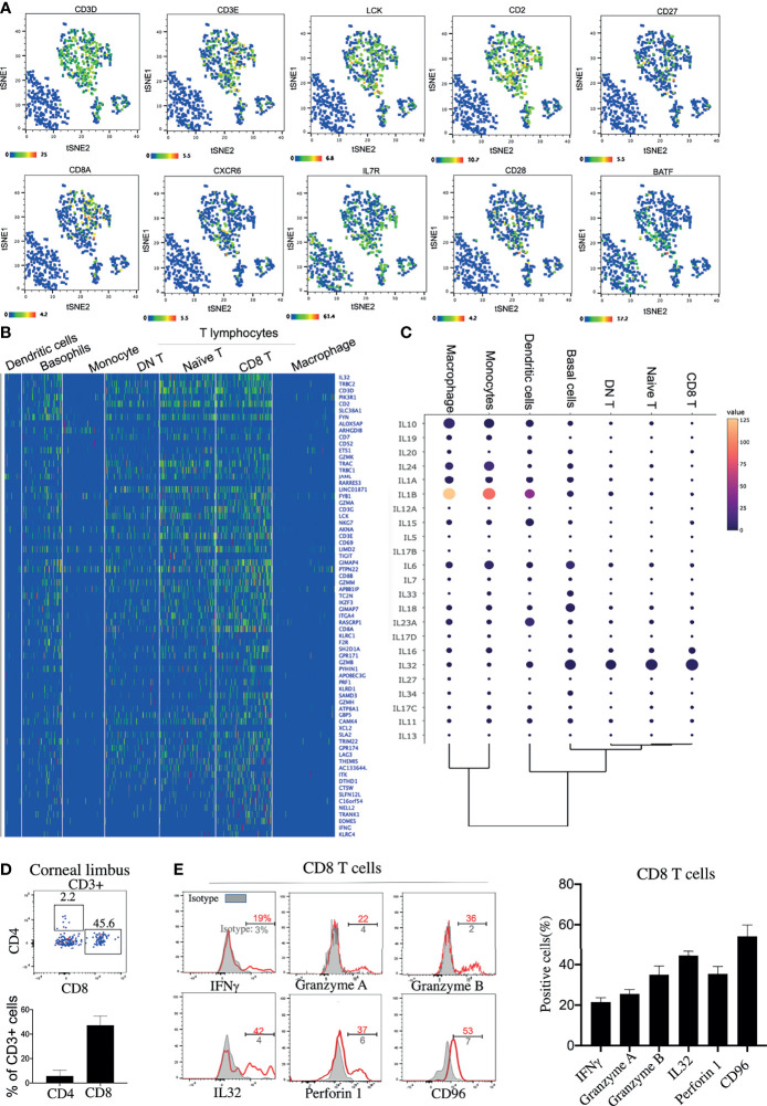
Background: The limbus is located at a 2-mm-wide area between the bulbar conjunctiva and the cornea and has been suggested to be the niche of corneal epithelial stem cells and immune cells. Like the skin and intestines, the cornea is also an important mucosal surface, and immune cells on the cornea play critical roles in immune surveillance to ensure barrier surface homeostasis and protection from various environmental damage and infections. Single-cell RNA sequencing (scRNA-seq) analysis of protein tyrosine phosphatase receptor type C positive (PTPRC+) hematopoietic cells from the corneal limbus could provide a single cell atlas of all the immune cell subsets.

Methods: We performed single-cell RNA sequencing to generate transcriptomic profile for 804 sort-purified hematopoietic cells from the corneal limbus of three healthy donors.

Results: Our analysis identified a primary transcriptomic pattern for multiple immune cell subtypes, including naive T cells, antiviral effector CD8+ T cells, and innate immune cells such as IDO1+ mature regulatory dendritic cells (mregDCs), macrophages, monocytes, and basophils in the human corneal limbus.

Conclusion: Overall, single-cell transcriptomic analysis of limbal immune cells suggested the possible contribution of these cells on the adaptive and innate immune response of the human cornea.

Keywords: MregDC; antiviral CD8+ T cells; chemotactic; corneal immune cells; single-cell transcriptome.


Conflict of interest statement

The authors declare that the research was conducted in the absence of any commercial or financial relationships that could be construed as a potential conflict of interest.

Figures

Figure 1
Figure 1
Identification of immune cell types in the cornea limbus. (A) Schematic of the single-cell RNA sequencing (scRNA-seq) workflow. The corneal limbus was gently digested to a single-cell suspension, enriched for hematopoietic cells by sorting PTPRC+ cells, and then scRNA-seq was performed. The data were then analyzed. (B) Gating strategy for sorting. Live CD45+ cells were selected. (C) Unbiased t-distributed stochastic neighbor embedding (t-SNE) clustering was used to determine the cell types and the frequency of the different immune cell types. (D) Heatmap of the specific marker genes for each cell type in scRNA-seq. (E) Network of enriched terms colored by cluster identity, where nodes that share the same cluster identity are typically close to each other. (F) Network of enriched terms colored by p-value, where terms containing more genes tend to have a more significant p-value.
Figure 2
Figure 2
Antiviral effector CD8+ T cells are a predominant lymphocyte subset on the cornea. (A) Feature t-distributed stochastic neighbor embedding (t-SNE) plot showing the expressions of marker genes enriched on corneal T cells. (B) Heatmap of the top expressed genes in the corneal T-cell subsets. (C) Dot plot showing the expressions of genes of the different cytokines (rows) on each cluster (columns). The color of each dot represents the average log-scaled expression of each gene across all cells of a given cluster. The size of the dot represents the fraction of cells in the cluster in which transcripts for that gene were detected. (D) Expressions of CD8 and CD4 proteins on corneal CD3+ T cells by flow cytometry (E) Expressions of selected cytokines and surface proteins on corneal CD8+ T cells by flow cytometry.
Figure 3
Figure 3
Immunoregulatory macrophages and monocytes recruit naive T cells by secreting CXCL16. (A) Heatmap of the top expressed genes in corneal macrophages, basophils, and monocytes. (B) Violin plot of the expressions of chemokine (C–X–C motif) ligands and chemokine (C–X–C motif) receptors in each cluster. (C) Expressions of IL-10, TGF-β, CXCL16, and CXCL12 on CD164+ corneal innate immune cells by flow cytometry. (D) Chemotactic activity of corneal CD3+ T cells to 100 ng/ml CXCL16 or the cell culture supernatant was determined with the Transwell migration system. (E) Pathway enrichment analysis of the differentially expressed genes in corneal macrophages, basophils, and monocytes. *Significant differences (P < 0.05, Student’s t test) from control groups.)
Figure 4
Figure 4
IDO1+ mature regulatory dendritic cells (mregDCs) are the major antigen-presenting cells on the cornea. (A) Dot plot showing the expressions of genes of the HLA subtypes (rows) in each cluster (columns). The color of each dot represents the average log-scaled expression of each gene across all cells of a given cluster. The size of the dot represents the fraction of cells in the cluster in which transcripts for that gene were detected. (B) Heatmap of the top expressed genes in corneal dendritic cells. (C) Pathway enrichment analysis of the differentially expressed genes in corneal dendritic cells. (D) The t-distributed stochastic neighbor embedding (t-SNE) plot showing comparative expressions of mregDC-specific signature genes from our data and those of Nakamizo et al. .

References

    1. Chan CC, Crowston JG, Tan R, Marin M, Charles S. Burden of Ocular Surface Disease in Patients With Glaucoma From Australia. Asia Pac J Ophthalmol (Phila) (2013) 2:79–87. doi: 10.1097/APO.0b013e31828372c2 - DOI - PubMed
    1. DelMonte DW, Kim T. Anatomy and Physiology of the Cornea. J Cataract Refract Surg (2011) 37:588–98. doi: 10.1016/j.jcrs.2010.12.037 - DOI - PubMed
    1. Akpek EK, Gottsch JD. Immune Defense at the Ocular Surface. Eye (Lond) (2003) 17:949–56. doi: 10.1038/sj.eye.6700617 - DOI - PubMed
    1. Reyes NJ, Saban DR. T Helper Subsets in Allergic Eye Disease. Curr Opin Allergy Clin Immunol (2014) 14:477–84. doi: 10.1097/ACI.0000000000000088 - DOI - PMC - PubMed
    1. Periman LM, Perez VL, Saban DR, Lin MC, Neri P. The Immunological Basis of Dry Eye Disease and Current Topical Treatment Options. J Ocul Pharmacol Ther (2020) 36:137–46. doi: 10.1089/jop.2019.0060 - DOI - PMC - PubMed

Publication types