. 2022 Feb 23:13:818017.
doi: 10.3389/fimmu.2022.818017. eCollection 2022.

STAT6 Blockade Abrogates Aspergillus-Induced Eosinophilic Chronic Rhinosinusitis and Asthma, A Model of Unified Airway Disease

Affiliations

STAT6 Blockade Abrogates Aspergillus-Induced Eosinophilic Chronic Rhinosinusitis and Asthma, A Model of Unified Airway Disease

Hua Sun et al. Front Immunol. .

Abstract

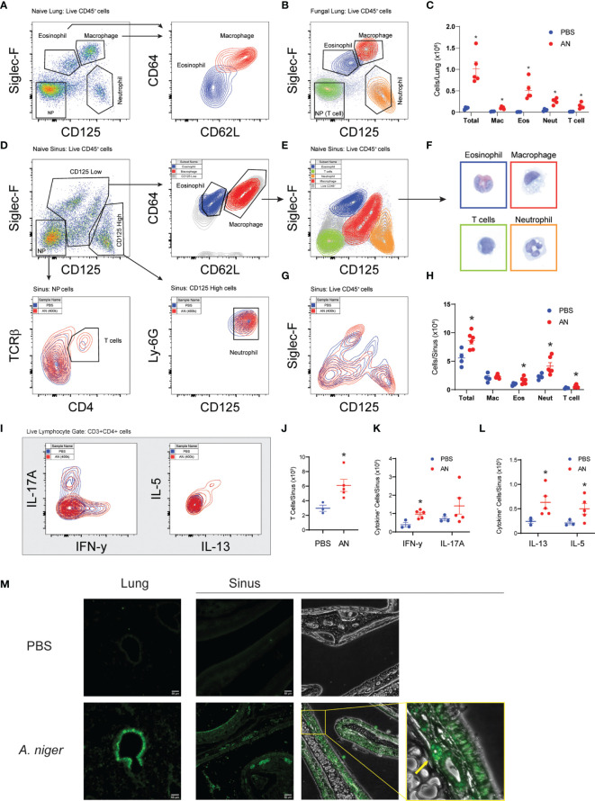
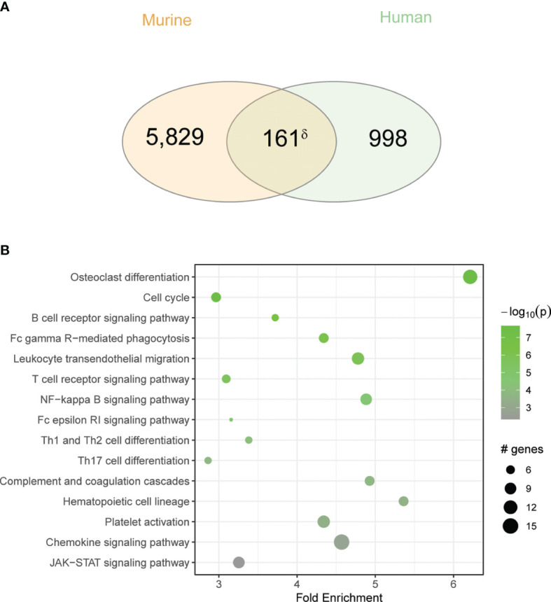
Unified airway disease, including concurrent asthma and chronic rhinosinusitis (CRS), is a common, but poorly understood disorder with no curative treatment options. To establish a murine model of chronic unified eosinophilic airway inflammation, mice were challenged with Aspergillus niger, and sinonasal mucosa and lung tissue were evaluated by immunohistochemistry, flow cytometry, and gene expression. Inhalation of A niger conidia resulted in a Th2-biased lung and sinus inflammation that typifies allergic asthma and CRS. Gene network and pathway analysis correlated with human disease with upregulation of not only the JAK-STAT and helper T-cell pathways, but also less expected pathways governing the spliceosome, osteoclast differentiation, and coagulation pathways. Utilizing a specific inhibitor and gene-deficient mice, we demonstrate that STAT6 is required for mycosis-induced sinus inflammation. These findings confirm the relevance of this new model and portend future studies that further extend our understanding of the immunopathologic basis of airway mycosis and unified airway disease.

Keywords: STAT-6; allergic; asthma; chronic rhinosinusitis; eosinophil; mouse model; mycosis; unified airway.


Conflict of interest statement

DC and JK hold intellectual property in PM-43I. The remaining authors declare that the research was conducted in the absence of any commercial or financial relationships that could be construed as a potential conflict of interest. The handling editor declared a shared affiliation with the author YZ at the time of review.

Figures

Figure 1
Figure 1
Intranasal fungal challenge promotes eosinophilic sinus inflammation. (A) Experimental outline of acute and chronic models. Mice were i.n. challenged with 4 × 10⁵ A. niger (AN) or PBS every other day for 2 weeks (acute) or 2–3 months (chronic) and assessed for inflammation. (B) Nasal airway lavage (NAL) inflammatory cells were quantified (C) from H&E-stained slides. (D) Representative H&E (left)- and PAS (right)-stained sinus sections from PBS- and AN-treated mice. Arrows indicate epithelial cells (green), eosinophils (red), neutrophils (blue), and mucus-stained goblet cells (orange). Results represent mean ± s.e.m. from two independent experiments; *p < 0.001, n ≥ 3 by two-way repeated measures ANOVA.
Figure 2
Figure 2
Airway mycosis induces eosinophilic, TH2-high inflammation in the upper and lower airways. Mice were challenged with 4 × 10⁵ A. niger (AN) or PBS daily for 2 months. Representative flow plots of lung single cell suspensions from (A) PBS or (B) fungal challenged (AN) wild-type C57BL/6 mice, with gates for eosinophils (Eos), macrophages (Mac), neutrophils (Neut), and TCRβ+CD4+ T cells in the negative population (NP), and sub-gates for Eos/Mac. (C) Differential cell counts from lungs. (D) Representative flow plots of naïve sinus, showing gating scheme for differential cell analysis and (E) sorting of cells for direct (F) imaging of H&E-stained cells (border colors match sorted population). (G) Overlay of fungal and PBS-treated mice and (H) differential cell counts of sinus inflammatory cells. (I) Representative flow plots of ex vivo stimulated TCRβ+CD4+ T cell cytokine production from sinuses was analyzed, with (J) total T cells, (K) IFN-γ+ and IL-17+, and (L) IL-13+ and IL-5+ cells quantified. (M) Fluorescent immunohistochemical staining of mouse sinus after PBS and A. niger challenge showing distribution of major basic protein, yellow box and arrow highlighting an eosinophil. Results presented as mean ± s.e.m. from at least two independent experiments with n = 3-5. *p < 0.05, n ≥ 3; by two-way ANOVA for differential counts and one-tailed t-test for cytokine production.
Figure 3
Figure 3
Fungal-mediated enrichment of inflammatory pathways of differentially expressed genes (DEGs) reveals variable gene isoform usage. Mice were i.n. challenged with A. niger (AN) or PBS as in Figure 1A, total RNA was isolated from sinus, and RNA-seq analysis was performed. (A) Volcano plot of DEGs in fungal challenged mouse sinus tissue vs. vehicle in which gene expression is depicted as fold change versus adjusted p-value (log10). (B) KEGG enrichment of significant pathways, with color indicating statistical significance and circle size indicating the number of genes. (C) Isoform switch analysis of fold change in sequenced gene isoforms for fungal challenged mice vs. naïve controls (dIF), with significantly switched isoforms shown in red. CD300lf (D–F) and Serpinb2 (G–I) sashimi plots of read coverage with isoform exon maps (D, G), total gene expression (E, H), and isoform usage (F, I). padj < 0.05 and q < 0.05; n = 3 by Student’s t-test. *p < 0.05; ***p < 0.001; ns, not significant.
Figure 4
Figure 4
Overlap between human CRS and chronic fungal challenged mouse sinus gene expression is enriched for inflammatory pathways. (A) Venn diagram showing the overlap between DEGs in the murine model to the CRS gene signature. (B) KEGG pathway enrichment analysis of overlapping genes. δp < 5e-10 by hypergeometric analysis.
Figure 5
Figure 5
Murine fungal rhinosinusitis has an asthmatic inflammatory gene signature. (A) Venn diagram showing overlap between DEGs and the asthma signature gene set for murine asthma21 and asthmatic human airway epithelial cells (hAEC). (B) KEGG pathway enrichment network and sub-networks including (C) Signal Transduction and (D) Immune System, for sinus DEGs (CRS; green), murine asthma (Asthma; blue), and co-enriched (Overlap; pink) pathways. (E) qPCR validation of select genes in co-enriched pathways (*) from sinus total RNA. δp < 5e-15 by hypergeometric analysis; padj < 0.05 for KEGG pathway enrichment; *p < 0.05, n = 3 by Student’s t-test.
Figure 6
Figure 6
Chronic airway mycosis promotes sinus epithelial cell alarmin, anti-microbial, and pro-asthma gene expression. (A) EpCam-positive (CD326+) epithelial cells were sorted from mice chronically challenged with 4 × 10⁵ A. niger (AN) or PBS every other day for 2 months for gene expression. (B) Diagram of relevant epithelial responses to fungal protease and Th2-driven inflammation. Protease activates protease activated receptor 2 (PAR-2) signaling to NF-κB and Erk1/2 to induce (C) alarmin and (D) antimicrobial gene expression. TH2 cytokines IL-13 and IL-4 signaling through IL-13Rα1 activate Erk1/2 and Stat6 to promote (E) pro-asthma gene expression. *p < 0.05, n = 3 by Student’s t-test.
Figure 7
Figure 7
STAT6 is essential for eosinophilic murine sinusitis. (A) Stat6-/- or C57BL/6 mice were challenged (i.n.) with 4 × 10⁵ A. niger (AN) or PBS for 3 months and lungs were assessed for inflammatory cell recruitment. (B) Representative flow analysis of Siglec-F and CD125 expression in wildtype (Wt) and Stat6-/- mice challenged with fungus (AN) or vehicle (PBS/DLPC) and (C) quantified. (D) Sinus inflammatory cell analysis from fungal challenged mice treated with 10 ng of PM-43i or vehicle (PBS/DLPC). *p < 0.05, n ≥ 3 by ANOVA. Data are from one of three representative experiments.

Similar articles

Cited by

References

    1. Orlandi RR, Kingdom TT, Hwang PH, Smith TL, Alt JA, Baroody FM, et al. . International Consensus Statement on Allergy and Rhinology: Rhinosinusitis. Int Forum Allergy Rhinol (2016) 6 Suppl 1:S22–209. doi: 10.1002/alr.21695 - DOI - PubMed
    1. Philpott CM, Erskine S, Hopkins C, Kumar N, Anari S, Kara N, et al. . Prevalence of Asthma, Aspirin Sensitivity and Allergy in Chronic Rhinosinusitis: Data From the UK National Chronic Rhinosinusitis Epidemiology Study. Respir Res (2018) 19:129. doi: 10.1186/s12931-018-0823-y - DOI - PMC - PubMed
    1. Bush RK, Prochnau JJ. Alternaria-Induced Asthma. J Allergy Clin Immunol (2004) 113:227–34. doi: 10.1016/j.jaci.2003.11.023 - DOI - PubMed
    1. Porter PC, Lim DJ, Maskatia ZK, Mak G, Tsai CL, Citardi MJ, et al. . Airway Surface Mycosis in Chronic TH2-Associated Airway Disease. J Allergy Clin Immunol (2014) 134:325–31. doi: 10.1016/j.jaci.2014.04.028 - DOI - PMC - PubMed
    1. Porter P, Susarla SC, Polikepahad S, Qian Y, Hampton J, Kiss A, et al. . Link Between Allergic Asthma and Airway Mucosal Infection Suggested by Proteinase-Secreting Household Fungi. Mucosal Immunol (2009) 2:504–17. doi: 10.1038/mi.2009.102 - DOI - PMC - PubMed

Publication types

Substances