Case Reports
. 2021 Sep-Dec;25(3):558.
doi: 10.4103/jomfp.jomfp_164_21. Epub 2022 Jan 11.

Granular cell ameloblastoma - A rare entity with recurrence after 48 years

Affiliations
Case Reports

Granular cell ameloblastoma - A rare entity with recurrence after 48 years

K R Anila et al. J Oral Maxillofac Pathol. 2021 Sep-Dec.

Abstract

A 64-year-old female with complaints of swelling right preauricular region was referred to our tertiary cancer center with fine-needle aspiration cytology (FNAC) report of mucoepidermoid carcinoma and radiological differential of malignant salivary gland neoplasm and sarcoma. On examination, there was a mass over her right parotid region Clinical diagnosis was malignant salivary gland neoplasm. Slide review of FNAC was inconclusive. Biopsy was done. Histopathology showed neoplasm comprising of nests of cells with abundant granular eosinophilic cytoplasm with focal area showing peripherally arranged columnar cells with palisading. On enquiry, the patient gave a history of surgery of right mandible 48 years back. Correlating histopathology and clinical history, a diagnosis of granular cell ameloblastoma was rendered. Radiological evaluation showed a solid-cystic lesion in the right masticator space. Right mandible showed only part of head of mandible consistent with previous surgery. Radical surgery was done. Final report confirmed the biopsy diagnosis.

Keywords: Diagnostic challenge; granular cell ameloblastoma; late recurrence.


Conflict of interest statement

There are no conflicts of interest.

Figures

Figure 1
Figure 1
Neoplasm comprising of nests of cells with abundant granular eosinophilic cytoplasm and peripherally placed nucleus. In focal areas at the periphery of the nests were seen columnar cells with palisading and reverse polarity (H&E, ×200)
Figure 2
Figure 2
Computed tomography showing solid-cystic lesion (asterisk) measuring 6.2 cm × 6 cm × 4.9 cm centered in the right masticator space. Right mandible ramus not visualized
Figure 3
Figure 3
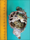
Solid cystic mass measuring 10 cm × 6 cm × 5 cm. Cut section gray-white with cystic area showing internal septations
Figure 4
Figure 4
(a) Neoplasm composed of nests of granular cells with abundant eosinophilic granular cytoplasm and peripherally compressed nucleus. Periphery showing the plexiform pattern of ameloblastoma (H&E, ×100). (b) The periphery of these nests of granular cells showed columnar cells with hyperchromatic, reversely polarized nuclei, arranged in a palisaded pattern (H&E, ×200)

Similar articles

References

    1. Taneeru S, Guttikonda VR, Yeluri S, Madala J. Granular cell ameloblastoma of jaw – Report of a case with an emphasis on its characterization. J Clin Exp Dent. 2013;5:e154–6. - PMC - PubMed
    1. Dave A, Arora M, Shetty VP, Saluja P. Granular cells in ameloblastoma: An enigma in diagnosis. Indian J Dent. 2015;6:211–4. - PMC - PubMed
    1. Nikitakis NG, Tzerbos F, Triantafyllou K, Papadimas C, Sklavounou A. Granular cell ameloblastoma: An unusual histological subtype report and review of literature. J Oral Maxillofac Res. 2011;1:e3. - PMC - PubMed
    1. Motahhary P, Etebarian A, Asareh F. Granular cell type of a unicystic ameloblastoma: An unusual case and review of the literature. J Oral Maxillofac Pathol. 2014;18:331. - PMC - PubMed
    1. Yamunadevi A, Madhushankari GS, Selvamani M, Basandi PS, Yoithapprabhunath TR, Ganapathy N. Granularity in granular cell ameloblastoma. J Pharm Bioallied Sci. 2014;6:S16–20. - PMC - PubMed

Publication types