Review
. 2022 Feb 25:16:738865.
doi: 10.3389/fnins.2022.738865. eCollection 2022.

More Than Meets the Eye: Art Engages the Social Brain

Affiliations
Review

More Than Meets the Eye: Art Engages the Social Brain

Janneke E P van Leeuwen et al. Front Neurosci. .

Abstract

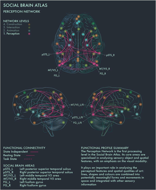
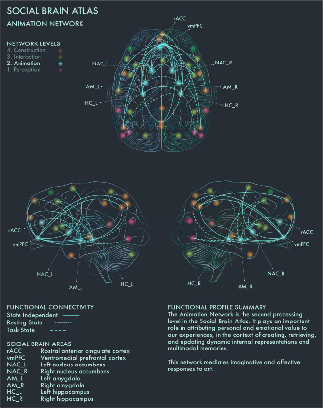
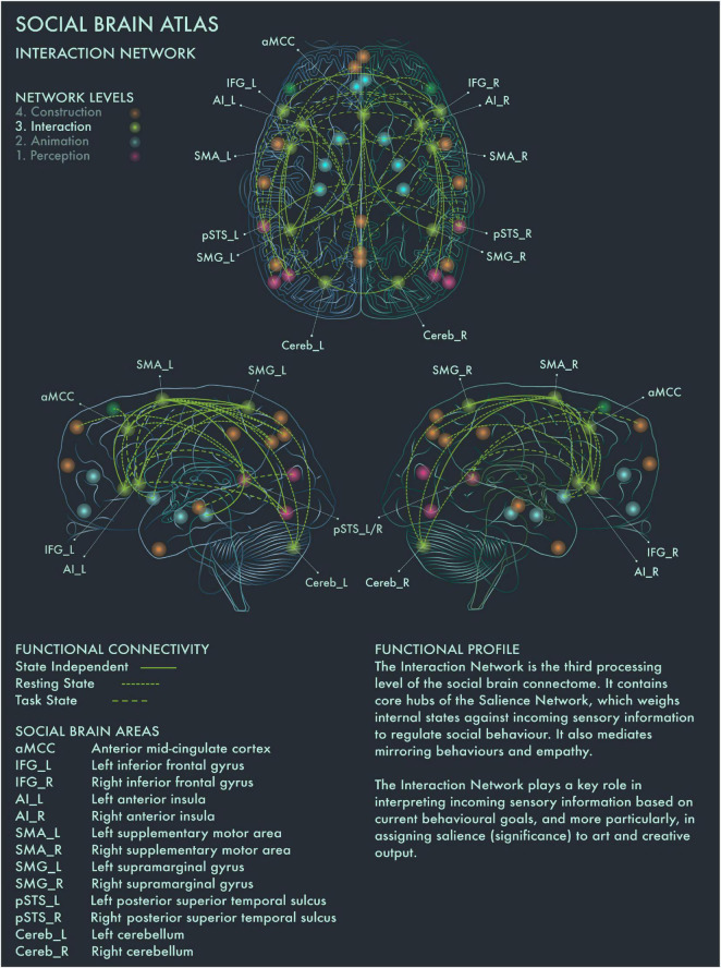
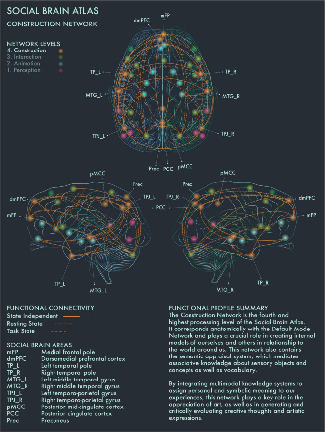
Here we present the viewpoint that art essentially engages the social brain, by demonstrating how art processing maps onto the social brain connectome-the most comprehensive diagram of the neural dynamics that regulate human social cognition to date. We start with a brief history of the rise of neuroaesthetics as the scientific study of art perception and appreciation, in relation to developments in contemporary art practice and theory during the same period. Building further on a growing awareness of the importance of social context in art production and appreciation, we then set out how art engages the social brain and outline candidate components of the "artistic brain connectome." We explain how our functional model for art as a social brain phenomenon may operate when engaging with artworks. We call for closer collaborations between the burgeoning field of neuroaesthetics and arts professionals, cultural institutions and diverse audiences in order to fully delineate and contextualize this model. Complementary to the unquestionable value of art for art's sake, we argue that its neural grounding in the social brain raises important practical implications for mental health, and the care of people living with dementia and other neurological conditions.

Keywords: art; art therapy; connectome; creativity; dementia; mental health; neuroaesthetics; social brain.


Conflict of interest statement

JL was the founder of the Thinking Eye, a social enterprise which translates novel insights from research into relationships between visual art processes and the social brain into services that aim to support psychological wellbeing and optimal cognitive functioning. The remaining authors declare that the research was conducted in the absence of any commercial or financial relationships that could be construed as a potential conflict of interest.

Figures

FIGURE 1
FIGURE 1
Social Brain Atlas Level 1: Perception Network. This figure shows the functional connectivity patterns of the Perception Network (PN), based on data derived from Alcalá-López et al. (2017) (see also Table 1(A)). The core nodes of the PN have been labeled as follows: Bilateral posterior superior temporal sulcus (pSTS_L/R); Bilateral middle temporal V5 area (MT/V5_L/R); Bilateral fusiform gyrus (FG_L/R). Areas beyond the PN to which these core nodes project are not labeled. Functional connections have been drawn as lines; a solid line indicates a functional connection independent of the brain state, a wide dotted line indicates a task-dependent functional connection and a narrow-dotted line indicates a functional connection during a brain state with no output task (the resting state). The PN is the first processing level in the social brain connectome Its core areas are specialized in analyzing sensory object and spatial features, with an emphasis on the visual modality. It plays an important role in analyzing the perceptual features and spatial qualities of art: lines, shapes and colors are combined into potentially meaningful forms and movements in space and integrated with other sensory information.
FIGURE 2
FIGURE 2
Social Brain Atlas Level 2: Animation Network. This figure shows the functional connectivity patterns of the Animation Network (AN), based on data derived from Alcalá-López et al. (2017) (see also Table 1(B)). Graphical conventions are the same as Figure 1. The core nodes of the AN have been labeled as follows: Rostral anterior cingulate cortex (rACC); Ventromedial prefrontal cortex (vmPFC); Bilateral nucleus accumbens (NAC_L/R); Bilateral amygdala (AM_L/R); Bilateral hippocampus (HC_L/R). The AN is the second processing level in the social brain connectome. It plays an important role in attributing personal and emotional value to our experiences, in the context of creating, retrieving, and updating dynamic internal representations and multimodal memories. This network mediates imaginative and affective responses to art.
FIGURE 3
FIGURE 3
Social Brain Atlas Level 3: Interaction Network. This figure shows the functional connectivity patterns of the Interaction Network (IN), based on data derived from Alcalá-López et al. (2017) (see also Table 1(C)). Graphical conventions are the same as Figure 1. The core nodes of the IN have been labeled as follows: Anterior mid-cingulate cortex (aMCC); Bilateral inferior frontal gyrus (IFG_L/R); Bilateral interior insula (AI_L/R); Bilateral supplementary motor area (SMA_L/R); Bilateral supramarginal gyrus (SMG_L/R; Bilateral posterior superior temporal sulcus (pSTS_L/R); Bilateral cerebellum (Cereb_L/R). The IN is the third processing level of the social brain connectome. It contains core hubs of the Salience Network, which weighs internal states against incoming sensory information to regulate social behavior. It also mediates mirroring behaviors and empathy. The IN plays a key role in interpreting incoming sensory information based on current behavioral goals, and more particularly, in assigning salience (significance) to art and creative output.
FIGURE 4
FIGURE 4
Social Brain Atlas Level 4: Construction Network. This figure shows the functional connectivity patterns of the Construction Network (CN), based on data derived from Alcalá-López et al. (2017) (see also Table 1(D)). Graphical conventions are the same as Figure 1. The core nodes of the CN have been labeled as follows: Medial frontal pole (mFP); Dorsomedial prefrontal cortex (dmPFC); Bilateral temporal pole (TP_L/R); Bilateral middle temporal gyrus (MTG_L/R); Bilateral temporo-parietal junction (TPJ_L/R); Posterior mid-cingulate cortex (pMCC); Posterior Cingulate Cortex (PCC); Precuneus (Prec). The CN is the fourth and highest processing level of the social brain connectome. It corresponds anatomically with the Default Mode Network and plays a crucial role in creating internal models of ourselves and others in relationship to the world around us. This network also contains the semantic appraisal system, which mediates associative knowledge about sensory objects and concepts as well as vocabulary. By integrating multimodal knowledge systems to assign personal and symbolic meaning to our experiences, the CN has a key role in the appreciation of art, as well as in generating and critically evaluating creative thoughts and artistic expressions.

References

    1. Abramović M. (1975). Art Must Be Beautiful, Artist Must Be Beautiful. Available Online at: https://www.moma.org/explore/inside_out/2010/04/06/listening-to-marina-a... [accessed January 6, 2021].
    1. Alcalá-López D., Smallwood J., Jefferies E., Van Overwalle F., Vogeley K., Mars R. B., et al. (2017). Computing the social brain connectome across systems and states. Cereb. Cortex 28 2207–2232. 10.1093/cercor/bhx121 - DOI - PubMed
    1. Alcalá-López D., Vogeley K., Binkofski F., Bzdok D. (2019). Building blocks of social cognition: mirror, mentalize, share? Cortex 118 4–18. 10.1016/j.cortex.2018.05.006 - DOI - PubMed
    1. Amodio D. M., Frith C. D. (2006). Meeting of minds: the medial frontal cortex and social cognition. Nat. Rev. Neurosci. 7 268–277. 10.1038/nrn1884 - DOI - PubMed
    1. Arnheim R. (1969). Visual Thinking/Rudolf Arnheim. Berkeley, CA: University of California Press.